Review
. 2020 Apr 30;40(4):BSR20193325.
doi: 10.1042/BSR20193325.

Current knowledge and recent advances in understanding metabolism of the model cyanobacterium Synechocystis sp. PCC 6803

Affiliations
Review

Current knowledge and recent advances in understanding metabolism of the model cyanobacterium Synechocystis sp. PCC 6803

Lauren A Mills et al. Biosci Rep. .

Abstract

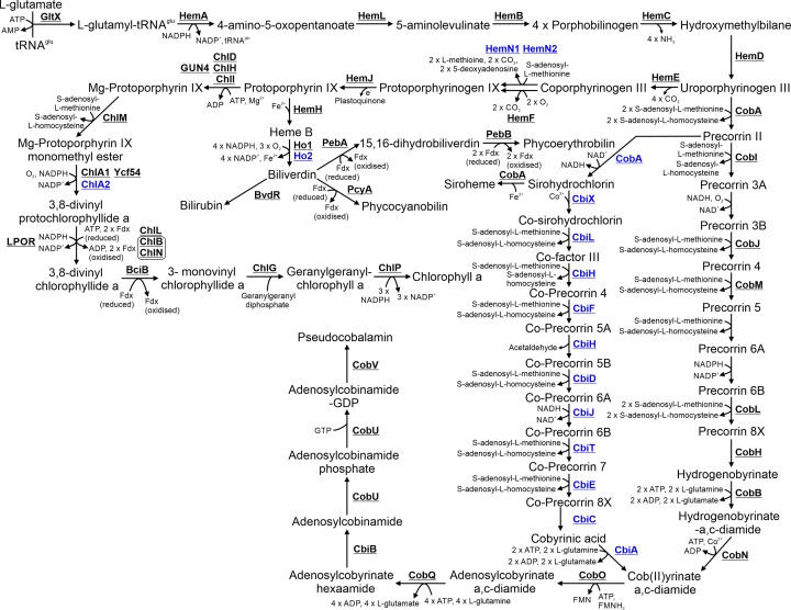
Cyanobacteria are key organisms in the global ecosystem, useful models for studying metabolic and physiological processes conserved in photosynthetic organisms, and potential renewable platforms for production of chemicals. Characterizing cyanobacterial metabolism and physiology is key to understanding their role in the environment and unlocking their potential for biotechnology applications. Many aspects of cyanobacterial biology differ from heterotrophic bacteria. For example, most cyanobacteria incorporate a series of internal thylakoid membranes where both oxygenic photosynthesis and respiration occur, while CO2 fixation takes place in specialized compartments termed carboxysomes. In this review, we provide a comprehensive summary of our knowledge on cyanobacterial physiology and the pathways in Synechocystis sp. PCC 6803 (Synechocystis) involved in biosynthesis of sugar-based metabolites, amino acids, nucleotides, lipids, cofactors, vitamins, isoprenoids, pigments and cell wall components, in addition to the proteins involved in metabolite transport. While some pathways are conserved between model cyanobacteria, such as Synechocystis, and model heterotrophic bacteria like Escherichia coli, many enzymes and/or pathways involved in the biosynthesis of key metabolites in cyanobacteria have not been completely characterized. These include pathways required for biosynthesis of chorismate and membrane lipids, nucleotides, several amino acids, vitamins and cofactors, and isoprenoids such as plastoquinone, carotenoids, and tocopherols. Moreover, our understanding of photorespiration, lipopolysaccharide assembly and transport, and degradation of lipids, sucrose, most vitamins and amino acids, and haem, is incomplete. We discuss tools that may aid our understanding of cyanobacterial metabolism, notably CyanoSource, a barcoded library of targeted Synechocystis mutants, which will significantly accelerate characterization of individual proteins.

Keywords: Synechocystis; comparative genomics; cyanobacteria; degradation; metabolism.


Conflict of interest statement

The authors declare that there are no competing interests associated with the manuscript.

Figures

Figure 1
Figure 1. Schematic detailing the ultrastructure of Synechocystis sp. PCC 6803 showing various subcellular components
Schematic adapted from [32,34].
Figure 2
Figure 2. Schematic detailing the pathways involved in central metabolism
Biosynthetic steps involved in glycolysis and gluconeogenesis are highlighted in red and blue respectively. Steps in the Entner–Doudoroff pathway are highlighted in green. Steps involved in the oxidative pentose phosphate pathway and the Calvin–Benson–Bassham cycle are highlighted in orange and purple, respectively. Fermentation pathways are highlighted in pink. Photorespiration pathways are highlighted in olive. Where enzymes catalyse reactions in two pathways, the arrows are split between their respective colours. The carboyxsome is represented as a purple octagon. Cofactors in each reaction are shown with the exception of protons, water, oxygen and inorganic phosphate.
Figure 3
Figure 3. Metabolism and degradation of nucleotide sugars and sugar osmolytes
Compounds highlighted in blue are substrates for lipopolysaccharide biosynthesis. Steps highlighted in grey are compounds and reactions not involved in these pathways but detailed in Figure 1. Cofactors in each reaction are shown with the exception of protons, water, oxygen and inorganic phosphate.
Figure 4
Figure 4. Metabolism of amino acids, cyanophycin, glutathione and iron–sulfur clusters
The 20 L-amino acids are highlighted in red while amino acids incorporated into peptidoglycan are highlighted in blue. The iron–sulfur biosynthetic pathways is highlighted in green. Steps highlighted in grey are compounds and reactions not involved in these pathways but detailed in Figure 1. Cofactors in each reaction are shown with the exception of protons, water, oxygen and inorganic phosphate.
Figure 5
Figure 5. Metabolism of nucleotides
The purine and pyrimidine biosynthesis pathways are highlighted in red and blue respectively. Possible nucleotide salvage pathways are highlighted in green. Cofactors in each reaction are shown with the exception of protons, water, oxygen and inorganic phosphate.
Figure 6
Figure 6. Metabolism of vitamins and cofactors
Detailed are the pathways for biosynthesis of (A) Biotin, (B) NAD+ and NADP+, (C) folate, (D) molybdenum cofactors, (E) riboflavin and FAD, (F) thiamine, (G) pantothenate and coenzyme (A and H) pyridoxal-5P. Vitamins and cofactors are highlighted in blue. Cofactors in each reaction are shown with the exception of protons, water, oxygen and inorganic phosphate.
Figure 7
Figure 7. Metabolism of membrane lipids, peptidoglycan and lipopolysaccharides
Membrane lipids are highlighted in blue. Steps highlighted in grey are compounds and reactions not involved in these pathways but detailed in Figure 2 and Figure 3. Cofactors in each reaction are shown with the exception of protons, water, oxygen and inorganic phosphate.
Figure 8
Figure 8. Metabolism of isoprenoids, quinols and carotenoids
Carotenoids are highlighted in blue. Cofactors in each reaction are shown with the exception of protons, water, oxygen and inorganic phosphate.
Figure 9
Figure 9. Metabolism of chlorophyll, phycobilin and pseudocobalamin
Proteins involved in anaerobic or low oxygen environment enzymatic steps are highlighted in blue. Cofactors in each reaction are shown with the exception of protons, water and inorganic phosphate.
Figure 10
Figure 10. Proteins involved in metabolite transport and conversion of nitrogen, sulfur and phosphate based compounds
Localization of transporters in either the PM or TM is detailed. Subunits in each complex may not all be membrane localized but soluble. Cofactors in each reaction are shown with the exception of protons, water, oxygen and inorganic phosphate.

References

    1. Rasmussen B., Fletcher I.R., Brocks J.J. and Kilburn M.R. (2008) Reassessing the first appearance of eukaryotes and cyanobacteria. Nature 455, 1101–U1109 10.1038/nature07381 - DOI - PubMed
    1. Holland H.D. (2006) The oxygenation of the atmosphere and oceans. Philosophical Transact. Royal Society B-Biological Sci. 361, 903–915 10.1098/rstb.2006.1838 - DOI - PMC - PubMed
    1. Zwirglmaier K. et al. . (2008) Global phylogeography of marine Synechococcus and Prochlorococcus reveals a distinct partitioning of lineages among oceanic biomes. Environ. Microbiol. 10, 147–161 - PubMed
    1. Galloway J.N. et al. . (2004) Nitrogen cycles: past, present, and future. Biogeochemistry 70, 153–226 10.1007/s10533-004-0370-0 - DOI
    1. Moisander P.H. et al. . (2017) Chasing after Non-cyanobacterial Nitrogen Fixation in Marine Pelagic Environments. Front. Microbiol. 8, 1736 10.3389/fmicb.2017.01736 - DOI - PMC - PubMed

Publication types

MeSH terms

Substances

LinkOut - more resources