. 2020 Jul;6(3):171-177.
doi: 10.1002/cjp2.160. Epub 2020 Mar 9.

Polymorphonuclear MDSCs are enriched in the stroma and expanded in metastases of prostate cancer

Affiliations

Polymorphonuclear MDSCs are enriched in the stroma and expanded in metastases of prostate cancer

Jiling Wen et al. J Pathol Clin Res. 2020 Jul.

Abstract

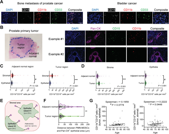
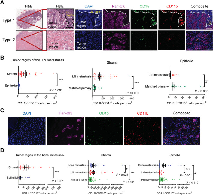
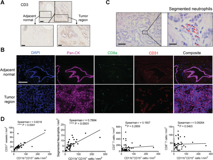
Myeloid-derived suppressor cells with polymorphonuclear morphology (PMN-MDSCs) contribute to the progression and immune evasion of prostate cancer. However, the spatial distribution of tumor-infiltrating PMN-MDSCs in primary and metastatic prostate cancer, especially in the context of comparison between the epithelial and stromal compartments of the tumor, has not been characterized. Here, we describe a multicolor immunofluorescence staining study of 90 primary tumors, 37 lymph node metastases (all with matched primary tumors) and 35 bone metastases using archived samples. CD11b+ CD15+ cells were identified as PMN-MDSCs and pan-cytokeratin+ cells were identified as prostate epithelial cells. We found that, in both primary tumor and metastases, PMN-MDSCs infiltrate much more readily in the stromal area compared with the epithelial area of the tumor regions. In comparison to the stromal area of primary tumors, the stromal area of either lymph node metastases or bone metastases was infiltrated with more PMN-MDSCs. In primary tumors, stromal PMN-MDSCs were associated with vascularization, segmented neutrophils, patient age and close juxtaposition to neoplastic epithelial cells. These results reveal the stroma rather than the epithelia of prostate cancer as the major hotbed for PMN-MDSCs and support the role of PMN-MDSCs in the metastatic progression of prostate cancer.

Keywords: bone metastasis; epithelial and stromal areas; lymph node metastasis; multicolor immunofluorescence staining; polymorphonuclear MDSC; prostate cancer; tumor microenvironment.


Figures

Figure 1
Figure 1
Increased infiltration of CD11b+ CD15+ PMN‐MDSCs in the stromal areas but not epithelial areas of primary prostate tumors. (A) Representative images of bone metastasis of PCa and bladder cancer showing the overlapping staining pattern of CD11b and CD33. Scale bar, 50 μm. (B) Representative image of a prostate tumor section with contours of the tumor and adjacent normal regions drawn by the pathologist, and two representative image sets of primary prostate tumors stained with the CD11b/CD15/pan‐CK/DAPI mIF panel. Scale bar, 100 μm. (C,D) Quantification results of CD11b+CD15+ PMN‐MDSCs. N = 90. Data represent mean ± standard error of the mean (SEM). P values calculated using a negative binomial regression model. (E) Illustration of the definition of epithelial area, stromal area and the distance between PMN‐MDSCs and nearest epithelial edge. (F) Violin plot comparing the distance between PMN‐MDSCs and the nearest epithelial area in the tumor and adjacent normal regions. **p = 0.0011 by Wilcoxon matched‐pair test. (G) Association of stroma‐residing PMN‐MDSCs, but not epithelia‐residing PMN‐MDSCs, in primary tumors with patient age (N = 90), tested by two‐tailed Spearman test.
Figure 2
Figure 2
Higher infiltration of PMN‐MDSCs in the stromal area of lymph node and bone metastases compared with the stromal area of primary tumors. (A) Representative images of the two types of lymph node metastasis samples recognized by the presence or absence of the lymph node/tumor interface in the H&E stain. Also shown are representative images of the lymph node metastasis samples stained with the CD11b/CD15/pan‐CK/DAPI mIF panel. Scale bar, H&E (left), 1000 μm; H&E (right) and IF images, 100 μm. (B) Quantification results of CD11b+CD15+ PMN‐MDSCs. N = 37 pairs of matched primary tumor and lymph node metastasis samples. (C) Representative images of the bone metastasis samples stained with the CD11b/CD15/pan‐CK/DAPI mIF panel. Scale bar, 100 μm. (D) Quantification results of CD11b+CD15+ PMN‐MDSCs. N = 35, 37 and 90 for bone metastases, lymph node metastases and primary tumors, respectively. In (B) and (D), data represent mean ± SEM. P values calculated using a negative binomial regression model.
Figure 3
Figure 3
Positive correlation of tumor stromal PMN‐MDSCs with vascularization and segmented neutrophils, evaluated using primary tumor samples (N = 46, SBMF). (A) Representative IHC images of CD3. Scale bar, 200 μm (top) and 100 μm (bottom). (B) Representative images of primary tumor stained with the CD31/CD8a/pan‐CK/DAPI mIF panel. Scale bar, 100 μm. (C) Representative images of visually recognized segmented neutrophils (red arrows). Scale bar, 100 μm (left) and 20 μm (right). (D) Correlation analysis of the numbers of tumor stromal PMN‐MDSCs with CD31+ vessels, segmented neutrophils, CD3+ total T cells, and CD8+ cytotoxic T cells, tested by two‐tailed Spearman test.

Similar articles

Cited by

References

    1. Veglia F, Perego M, Gabrilovich D. Myeloid‐derived suppressor cells coming of age. Nat Immunol 2018; 19: 108–119. - PMC - PubMed
    1. Gabrilovich DI. Myeloid‐derived suppressor cells. Cancer Immunol Res 2017; 5: 3–8. - PMC - PubMed
    1. Torre LA, Bray F, Siegel RL, et al Global cancer statistics, 2012. CA Cancer J Clin 2015; 65: 87–108. - PubMed
    1. Lu X, Horner JW, Paul E, et al Effective combinatorial immunotherapy for castration‐resistant prostate cancer. Nature 2017; 543: 728–732. - PMC - PubMed
    1. Hossain DM, Pal SK, Moreira D, et al TLR9‐targeted STAT3 silencing abrogates immunosuppressive activity of myeloid‐derived suppressor cells from prostate cancer patients. Clin Cancer Res 2015; 21: 3771–3782. - PMC - PubMed

Publication types