. 2020 Apr 14;145(8):3090-3099.
doi: 10.1039/d0an00097c.

Evaluation of top-down mass spectrometry and ion-mobility spectroscopy as a means of mapping protein-binding motifs within heparin chains

Affiliations

Evaluation of top-down mass spectrometry and ion-mobility spectroscopy as a means of mapping protein-binding motifs within heparin chains

Yunlong Zhao et al. Analyst. .

Abstract

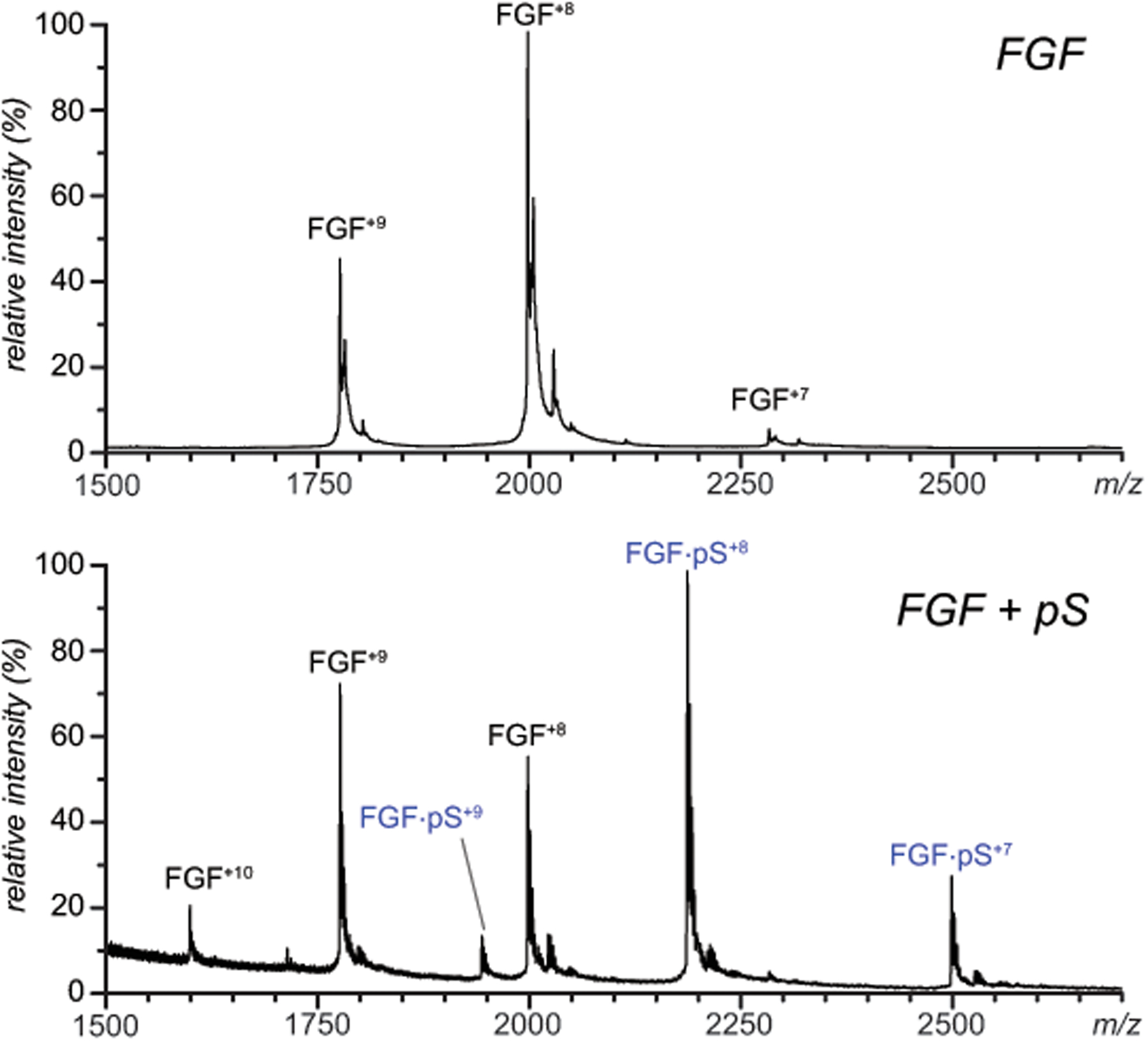
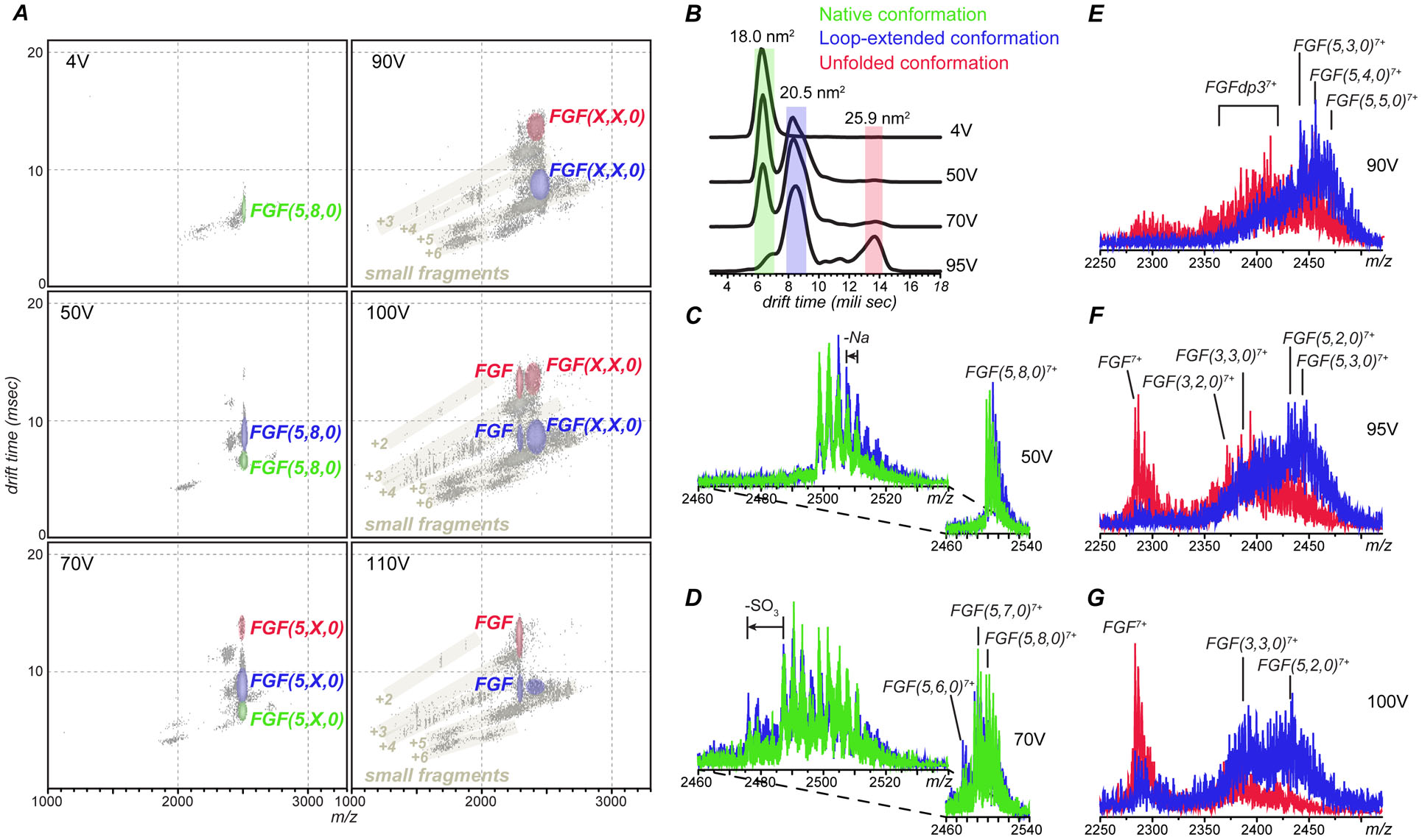
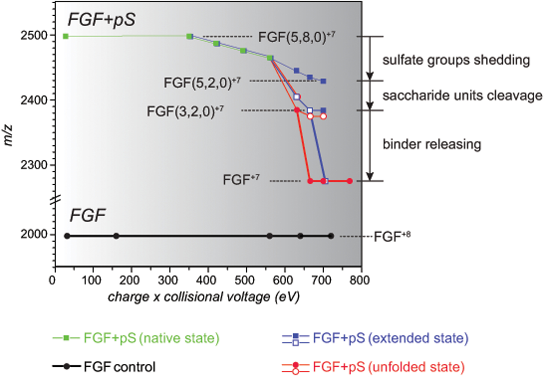
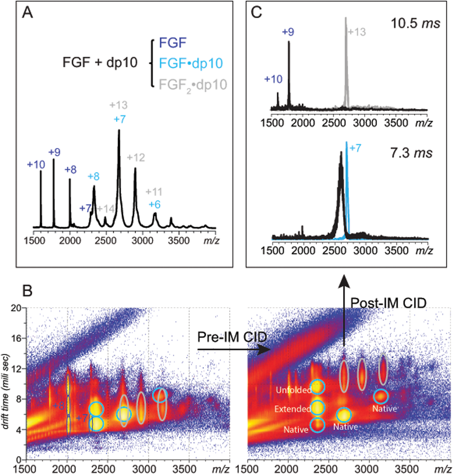
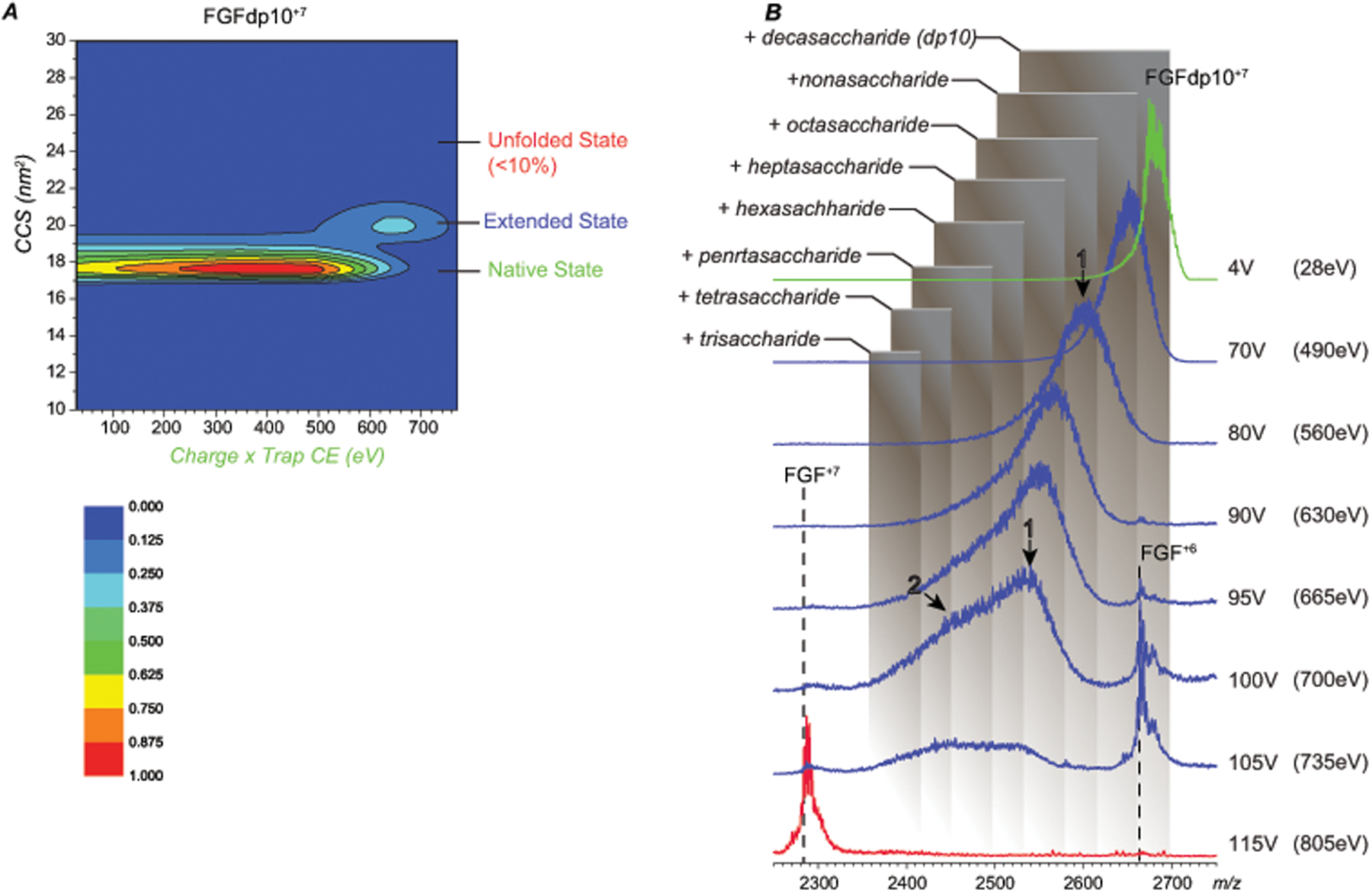
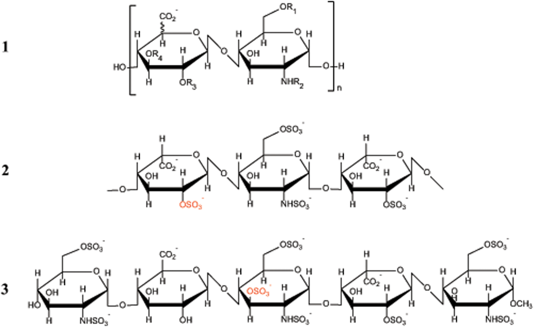
Identifying structural elements within heparin (as well as other glycosaminoglycan) chains that enable their interaction with a specific client protein remains a challenging task due to the high degree of both intra- and inter-chain heterogeneity exhibited by this polysaccharide. The new experimental approach explored in this work is based on the assumption that the heparin chain segments bound to the protein surface will be less prone to collision-induced dissociation (CID) in the gas phase compared to the chain regions that are not involved in binding. Facile removal of the unbound chain segments from the protein/heparin complex should allow the length and the number of sulfate groups within the protein-binding segment of the heparin chain to be determined by measuring the mass of the truncated heparin chain that remains bound to the protein. Conformational integrity of the heparin-binding interface on the protein surface in the course of CID is ensured by monitoring the evolution of collisional cross-section (CCS) of the protein/heparin complexes as a function of collisional energy. A dramatic increase in CCS signals the occurrence of large-scale conformational changes within the protein and identifies the energy threshold, beyond which relevant information on the protein-binding segments of heparin chains is unlikely to be obtained. Testing this approach using a 1 : 1 complex formed by a recombinant form of an acidic fibroblast growth factor (FGF-1) and a synthetic pentasaccharide GlcNS,6S-GlcA-GlcNS,3S,6S-IdoA2S-GlcNS,6S-Me as a model system indicated that a tri-saccharide fragment is the minimal-length FGF-binding segment. Extension of this approach to a decameric heparin chain (dp10) allowed meaningful binding data to be obtained for a 1 : 1 protein/dp10 complex, while the ions representing the higher stoichiometry complex (2 : 1) underwent dissociation via asymmetric charge partitioning without generating truncated heparin chains that remain bound to the protein.


Figures

Figure 1.
Figure 1.
Native MS of a 5 μM solution of FGF-1 (100 mM ammonium acetate) obtained from heparin-free solution (blue trace) and in the presence of 50 μM synthetic pentasaccharide, fondaparinux (red). The synthetic pentasaccharide is annotated on the graph as pS.
Figure 2.
Figure 2.
The effect of collisional energy on population of fragment ions derived from dissociation of FGF/pentasaccharide complexes (charge state +7) and their gas-phase mobility represented by (A) two-dimensional IMS-MS plots, (B) extracted CIU profile of the precursor ion and (C-G) conformer-specific fragment ion spectra where the fragment ions are derived from compact, near-native (green), partially unfolded (blue) and fully unfolded (red) precursor ions at different activation energies. The three numbers in parentheses are used to designate composition of GAG fragments (the number of saccharide monomers in the chain, the number of sulfate groups and the number of acetyl groups) following the commonly accepted convention.
Figure 3.
Figure 3.
Mass evolution of FGF·pS complexes as a function of collisional energy plotted for three different precursor conformations: native (green), partially unfolded (blue) and fully unfolded (red). The boldness of line indicates the category of mass change: sulfate shedding (thin), saccharide unit(s) removal (normal) and complete ligand release (bold). Only the lowest-m/z edge of the ionic distributions is represented on plot. The shaded squares/circles represent the dominant features in the mass spectra (relative abundance ≥ 45%); the minor features are represented with the open squares/circles.
Figure 4.
Figure 4.
Native MS/IMS characterization of SEC-purified FGF/dp10 complexes. A: a mass spectrum of the FGF/dp10 in 100 mM ammonium acetate; the charge states are labeled for the heparin-free protein (blue), as well as for 1:1 (cyan) and 2:1 (gray) FGF/dp10 complexes. B (left panel): a two-dimensional IMS/MS plot of for the mass spectrum shown in (A) showing significant overlap of ions representing FGF/dp10 complexes with different stoichiometries (cyan and gray ovals) in both m/z and drift time domains (left). B (right panel): modest pre-IM collisional activation (10 V) allows the ions representing the two different FGF/dp10 complexes to be completely separated. C: the pre-IM separated complexes can be activated independently post-IM, giving rise to distinct fragmentation patterns at collisional activation of 80 V.
Figure 5.
Figure 5.
CCS (A) and m/z (B) evolution of ions representing the 1:1 FGF/dp10 complex (charge state +7) following collisional activation. The shaded boxes in panel B represent m/z ranges for FGF bound to truncated forms of the heparin oligomer of varying lengths.
Scheme 1.
Scheme 1.
Chemical structures of the repeat units of heparin (1); the FGF-1 binding trisaccharide of heparin (2) and the synthetic antithrombotic fondaparinux (3). The typical substitution patterns in 1 are as follows: R1, R3 = H or SO3‾; R2 = H, C(O)CH3 or SO3‾; and R4 = H (common) or SO3‾ (rare).

References

    1. Pomin VH and Mulloy B, Curr. Opin. Struct. Biol, 2015, 34, 17–25. - PubMed
    1. Jin L, Abrahams JP, Skinner R, Petitou M, Pike RN and Carrell RW, Proceedings of the National Academy of Sciences of the United States of America, 1997, 94, 14683–14688. - PMC - PubMed
    1. Weitz JI, The New England journal of medicine, 1997, 337, 657–662. - PubMed
    1. McNeely TB and Griffith MJ, Blood, 1985, 65, 1226–1231. - PubMed
    1. Lindahl U, Haemostasis, 1999, 29 Suppl S1, 38–47. - PubMed