Comparative Study
. 2020 Mar 9;61(3):5.
doi: 10.1167/iovs.61.3.5.

Comparison of Neuroprotective Effects of Monomethylfumarate to the Sigma 1 Receptor Ligand (+)-Pentazocine in a Murine Model of Retinitis Pigmentosa

Affiliations
Comparative Study

Comparison of Neuroprotective Effects of Monomethylfumarate to the Sigma 1 Receptor Ligand (+)-Pentazocine in a Murine Model of Retinitis Pigmentosa

Haiyan Xiao et al. Invest Ophthalmol Vis Sci. .

Abstract

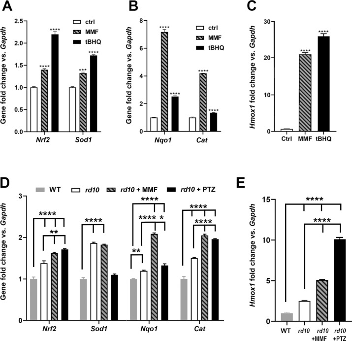
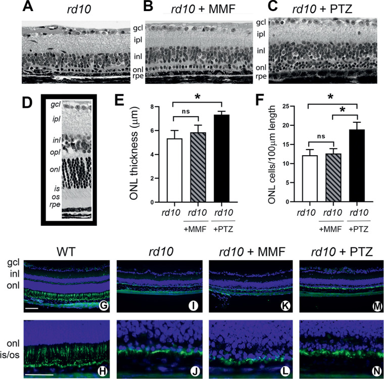
Purpose: Activating the cell survival modulator sigma 1 receptor (Sig1R) delays cone photoreceptor cell loss in Pde6βrd10/J (rd10) mice, a model of retinitis pigmentosa. Beneficial effects are abrogated in rd10 mice lacking NRF2, implicating NRF2 as essential to Sig1R-mediated cone neuroprotection. Here we asked whether activation of NRF2 alone is sufficient to rescue cones in rd10 mice.

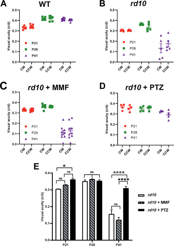
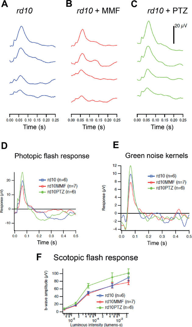
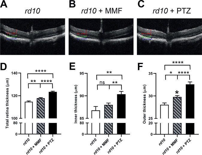
Methods: Expression of antioxidant genes was evaluated in 661W cells and in mouse retinas after treatment with monomethylfumarate (MMF), a potent NRF2 activator. Rd10 mice were administered MMF (50 mg/kg) or the Sig1R ligand (+)-pentazocine (PTZ; 0.5 mg/kg) intraperitoneally (every other day, P14-42). Mice were evaluated for visual acuity (optokinetic tracking response), retinal function (electroretinography) and architecture (SD-OCT); histologic retinal sections were evaluated morphometrically.

Results: MMF treatment increased Nrf2, Nqo1, Cat, Sod1, and Hmox1 expression in vitro and in vivo. Visual acuity of (+)-PTZ-treated rd10 mice was similar to wild-type mice; however, MMF treatment did not alter acuity compared with nontreated rd10 mice. Cone electroretinography b-wave amplitudes were greater in PTZ-treated than nontreated or MMF-treated rd10 mice. SD-OCT assessment of retinal thickness was greater in (+)-PTZ-treated mice versus nontreated or MMF-treated rd10 mice. Morphometric assessment of the outer nuclear layer revealed approximately 18 cells/100 µm retinal length in (+)-PTZ-treated rd10 mice, but only approximately 10 to 12 cells/100 µm in MMF-treated and nontreated rd10 retinas.

Conclusions: Activation of NRF2 using MMF, at least at our dosing regimen, is insufficient to attenuate catastrophic photoreceptor damage characteristic of rd10 mice. The data prompt investigation of additional mechanisms involved in Sig1R-mediated retinal neuroprotection.


Conflict of interest statement

Disclosure: H. Xiao, None; J. Wang, None; A. Saul, None; S.B. Smith, None

Figures

Figure 1.
Figure 1.
Evaluation of antioxidant gene expression in 661W cells treated with MMF. The 661W cells were incubated with MMF [400 µM] for 8 hours. RNA was isolated and subjected to qRT-PCR to analyze expression of (A) Nrf2, Sod1, (B) Nqo1, Cat, and (C) Hmox1. Primer pairs used for analysis are listed in Table 1. Data are mean ± SEM of three independent assays. Significant differences (***P < 0.001; ****P < 0.0001) are in reference to the control (nontreated cells). The positive control used to induce Nrf2 was tBHQ (20 µM). RNA was isolated from neural retinas of WT, rd10, rd10+MMF, rd10+PTZ mice and subjected to qRT-PCR analysis of (D) Nrf2, Sod1, Nqo1, Cat, and (E) Hmox1 using the primer pairs listed in Table 1. Data are the mean ± SEM of three to four assays. Significantly different from gene expression in WT mice (**P < 0.01; ****P < 0.0001).
Figure 2.
Figure 2.
Assessment of visual acuity in rd10 mice treated with MMF compared with nontreated and PTZ-treated. The rd10 mice were administered MMF or (+)-PTZ every other day beginning at P14 and were compared with nontreated rd10 mice. The optokinetic tracking response (OKR) was measured using the OptoMotry system to assess visual acuity at P21, P28, and P41. Data are expressed as cycles/degree (c/d) for (A) WT mice, (B) nontreated rd10 mice, (C) rd10+MMF mice, and (D) rd10 +PTZ mice. The data were summarized for comparison (E). Significance is depicted as *P < 0.05, ****P < 0.0001. CW, left eye; CCW, right eye; ns, not significant.
Figure 3.
Figure 3.
Assessment of retinal function by photopic and scotopic ERG in rd10 mice treated with MMF compared with nontreated and PTZ-treated mice. Rd10 mice were administered MMF or (+)-PTZ every other day beginning at P14 and were compared with nontreated rd10 mice. Photopic and scotopic ERG responses were performed on rd10 mice, rd10+MMF mice, and rd10+PTZ mice at P35. Averaged photopic responses to 5ms flashes at a series of intensities were provided for (A) rd10, (B) rd10+MMF, and (C) rd10+PTZ mice at P35. (D) Averaged responses to the highest intensity photopic flashes. (E) Averaged kernels derived from responses to natural noise stimuli. (F) Averaged scotopic ERG responses to 5-ms flashes at a series of intensities for rd10, rd10+MMF and rd10+PTZ. Intensities were in units of candela-seconds per meter squared.
Figure 4.
Figure 4.
Assessment of retinal structure in vivo using SD-OCT in rd10 mice treated with MMF compared with nontreated and PTZ-treated mice. Rd10 mice were administered MMF or (+)-PTZ every other day beginning at P14 and were compared with nontreated rd10 mice. SD-OCT was performed on mice at P42. Representative SD-OCT images are shown for (A) rd10 (nontreated) mice, (B) rd10+MMF mice, and (C) rd10+PTZ mice. Data from segmentation analysis for (D) total retinal thickness, (E) thickness of the inner retina, and (F) thickness of the outer retina. Significance is depicted as *P < 0.05; **P < 0.01; ****P < 0.0001. ns, not significant. Data are the mean ± SEM of analyses in 5 to 15 mice per group.
Figure 5.
Figure 5.
Assessment of retinal histologic structure and detection of cone photoreceptor cells in rd10 mice treated with MMF compared with nontreated and PTZ-treated. Rd10 mice were administered MMF or (+)-PTZ every other day beginning at P14 and were compared with nontreated rd10 mice. After the functional tests, mice were euthanized at P42 and eyes taken for plastic embedding or frozen sections. The plastic sections were stained with hematoxylin and eosin and viewed by light microscopy. Representative retinas are provided for (A) rd10 (nontreated), (B) rd10 + MMF, (C) rd10+PTZ. (D) A representative retinal section of an age-matched WT mouse. Morphometric analysis was performed on the retinal sections; data are presented as mean ± SEM for (None;) the measurement of ONL thickness and (F) the number of photoreceptor cell nuclei in the ONL expressed per 100 µm retinal length. Significance *P < 0.05. ns, not significant. Frozen sections were used for immunodetection of cone photoreceptor cells using FITC-peanut agglutinin (PNA) shown at low and high magnification, respectively for WT (G, H), rd10-nontreated (I, J), rd10 + MMF (K, L), and rd10+PTZ (M, N). Green fluorescence is associated with FITC-PNA; nuclei fluoresce blue owing to labeling using 4′,6-diamidino-2-phenylindole (DAPI) staining. gcl, ganglion cell layer; ipl, inner plexiform layer; inl, inner nuclear layer; opl, outer plexiform layer; onl, outer nuclear layer; is, inner segment; os, outer segment. Calibration bar: 100 µm.

Similar articles

Cited by

References

    1. Duncan JL, Pierce EA, Laster AM, Daiger SP, Birch DG, Ash JD, et al. ., the Foundation Fighting Blindness Scientific Advisory Board. Inherited retinal degenerations: current landscape and knowledge gaps. Transl Vis Sci Technol. 2018; 7: 6. - PMC - PubMed
    1. Daiger SP, Sullivan LS, Bowne SJ. Genes and mutations causing retinitis pigmentosa. Clin Genet. 2013; 84: 132–141. - PMC - PubMed
    1. Hernández-Rabaza V, López-Pedrajas R, Almansa I. Progesterone, lipoic acid, and sulforaphane as promising antioxidants for retinal diseases: a review. Antioxidants (Basel). 2019; 8(3). - PMC - PubMed
    1. Athanasiou D, Aguila M, Bellingham J, Li W, McCulley C, Reeves PJ, et al. .. The molecular and cellular basis of rhodopsin retinitis pigmentosa reveals potential strategies for therapy. Prog Retin Eye Res. 2018; 62: 1–23. - PMC - PubMed
    1. Campochiaro PA, Mir TA. The mechanism of cone cell death in retinitis pigmentosa. Prog Retin Eye Res. 2018; 62: 24–37. - PubMed

Publication types

MeSH terms