. 2020 Mar 9;61(3):7.
doi: 10.1167/iovs.61.3.7.

Cd9 Protects Photoreceptors from Injury and Potentiates Edn2 Expression

Affiliations

Cd9 Protects Photoreceptors from Injury and Potentiates Edn2 Expression

Toshiro Iwagawa et al. Invest Ophthalmol Vis Sci. .

Abstract

Purpose: Cd9 is a tetraspanin membrane protein that plays various roles in tissue development and disease pathogenesis, especially in cancer, but the expression patterns and function of Cd9 in retinal development and disease are not well understood. We asked its roles during retinal photoreceptor degeneration by using CD9-knockout mice.

Methods: Cd9 knockout mice and rd1 mice were used to examine roles of Cd9 for progression of photoreceptor degeneration. Reverse transcription-polymerase chain reaction and immunohistochemistry were mainly used as analytical methods.

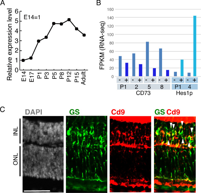
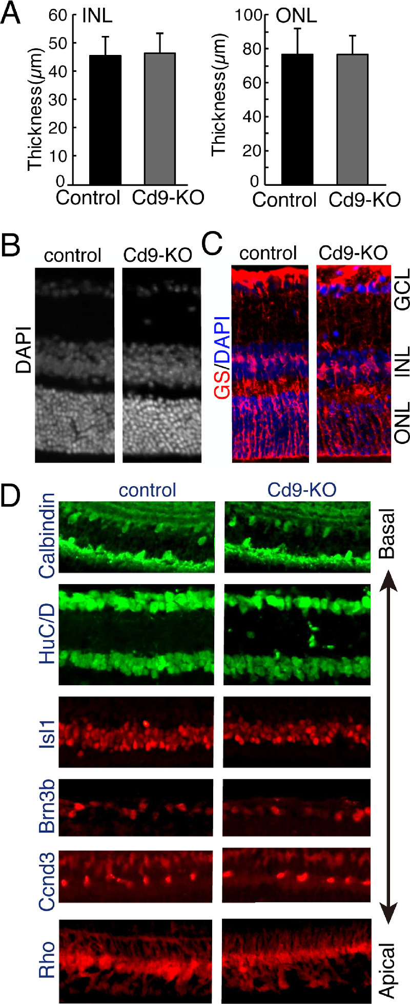
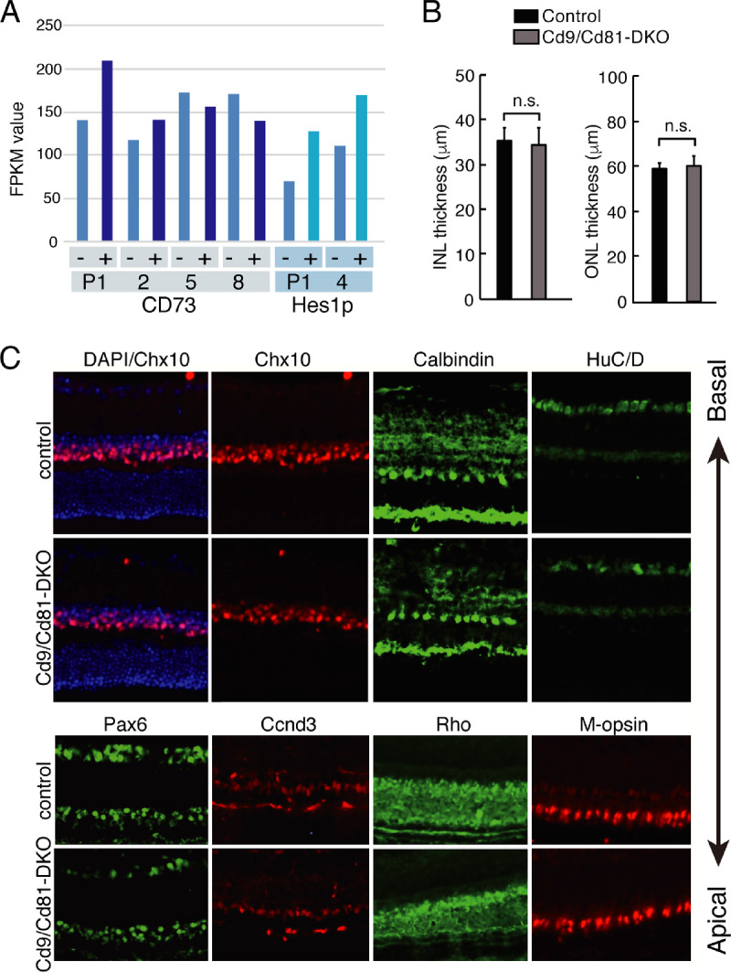
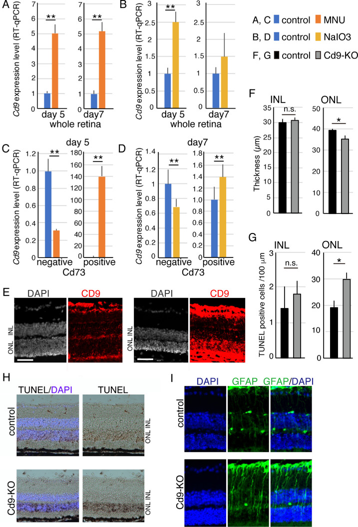
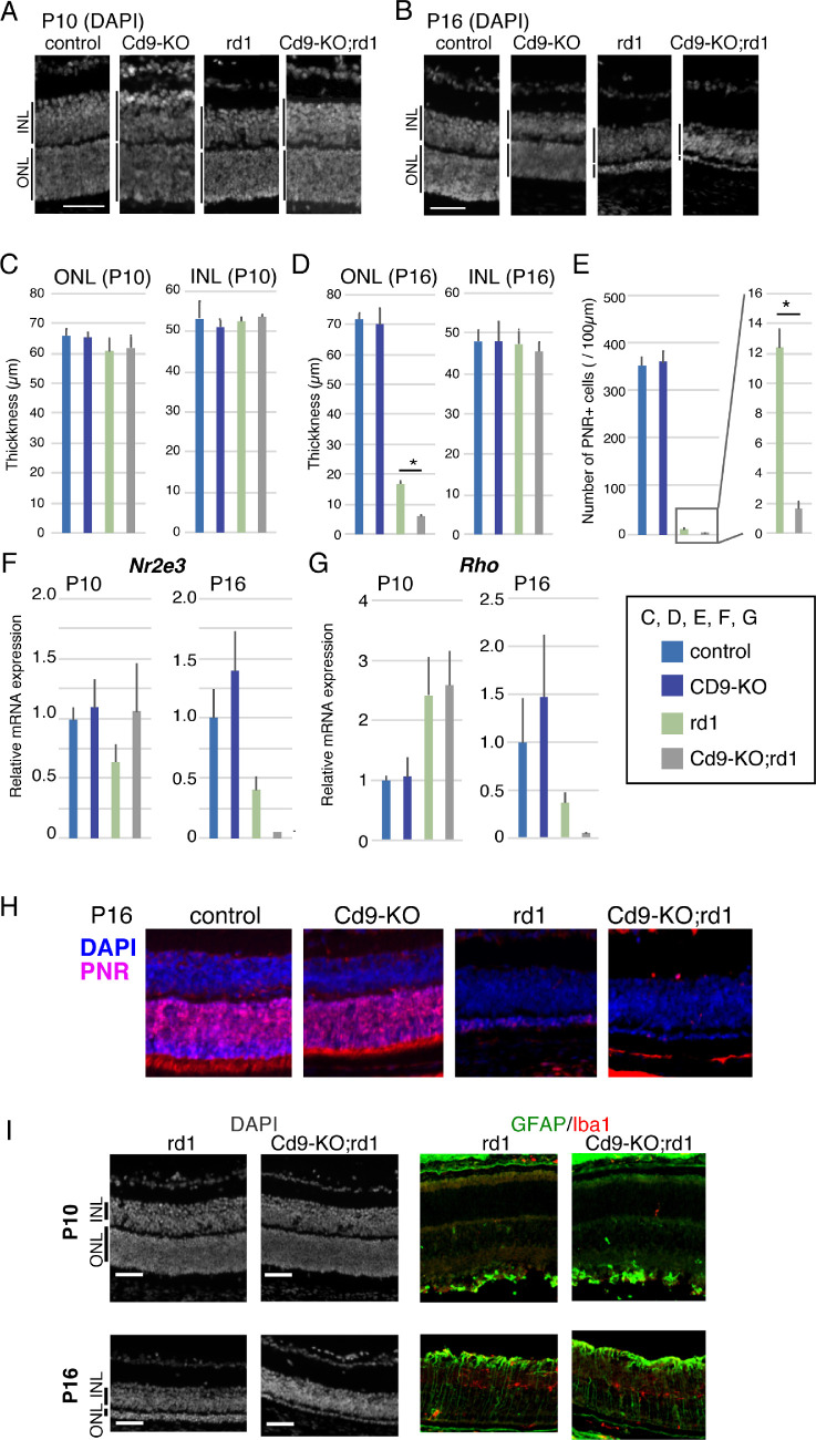
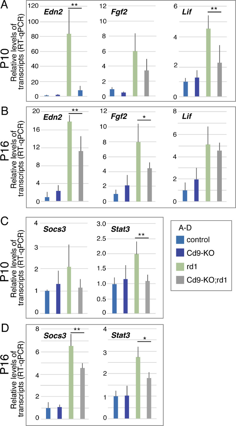
Results: Cd9 transcripts were only weakly expressed in retina at embryonic day 14, but its expression level subsequently increased and peaked at around postnatal day 12. In 6-week-old female mice derived retina, mRNA expression decreased slightly but was maintained at a significant level. Published RNA-sequencing data and immunohistochemistry indicated that Cd9 was expressed abundantly in Müller glia and weakly in other retinal neurons. Notably, when photoreceptors were damaged, Cd9 expression was increased in rod photoreceptors and decreased in Müller glia. Cd9 knockout mice retinas developed normally; however, once the retina suffered damage, degeneration of photoreceptors was more severe in Cd9 knockout retinas than control retinas. Induction of Edn2, which is known to protect against photoreceptor damage, was severely hampered. In addition, induction of Socs3, which is downstream of gp130 (Il6st), was weaker in Cd9 knockout retinas.

Conclusions: Taken together, these findings indicate that, although Cd9 was dispensable for normal gross morphological development, it protected rod photoreceptors and enhanced Edn2 expression, possibly through modulation of gp130 signaling.


Conflict of interest statement

Disclosure: T. Iwagawa, None; Y. Aihara, None; D. Umutoni, None; Y. Baba, None; A. Murakami, None; K. Miyado, None; S. Watanabe, None

Figures

Figure 1.
Figure 1.
Transition of the expression of Cd9 during retinal development. (A) Transition of expression levels of Cd9 transcripts during retinal development. Whole retinas at indicated developmental stage were harvested, and RT-qPCR was done using a primer specific for Cd9. The values were normalized to Cq value of Gapdh and Sdha, and expressed as relative to E14 value. An 8-week-old mouse was analyzed as an adult mouse. (B) Expression levels of Cd9 transcripts in subpopulations of developing retina. Fragments per kilobase of exon per million mapped fragments (FPKM) value of RNA-seq data of purified Cd73 positive rod photoreceptors and Cd73 negative other cells at P1, 2, 5, 8 (GEO: GSE71462), and Hes1-promoter positive Müller glia and negative other cells at P1 and P4 (GEO: GSE86199) are shown. (C) Immunohistochemistry using anti-Cd9 and -GS, which marks Müller glia, antibodies in a P10 mouse retinal frozen section. One representative sample out of samples from three independent mice was shown. Nuclei were visualized by staining with DAPI (gray). Arrowheads indicate GS and Cd9 double positive cells. Scale bar: 50 µm.
Figure 2.
Figure 2.
The retina develops appropriately in Cd9-KO mouse. (A) The thickness of INL and ONL of the retinas of adult control or Cd9-KO mice at 8 weeks was measured in frozen sectioned retina. The values are average of more than 9 sections from 3 mice for each genotype with standard deviation. (B-D) Immunohistochemistry by using indicated antibodies of frozen sections of the retinas of control or Cd9-KO mice was performed. All panels are shown as ONL to the bottom. The antibodies label specific subsets of retinal cells; GS (Müller glia), Calbindin (subsets of amacrine and horizontal), HuC/D (amacrine and RGC), Isl1 (bipolar), Brn3b (RGC), Ccnd3 (Müller glia), Rho (rod photoreceptor). Nuclei were visualized by staining with DAPI in B (gray) and C (blue).
Figure 3.
Figure 3.
The retinas of Cd9 and Cd81 double knockout mice (Cd9/Cd81-DKO) showed comparable morphological development as in control retina. (A) Expression levels of Cd81 transcript in developing retina. FPKM values of Cd81 from RNA-seq of Cd73-positive rod photoreceptors and Cd73-negative other cells at P1, 2, 5, 8, and Hes1-promoter (Hes1p)-positive Müller glia and -negative other cells at P1 and P4 are shown. (B) Thickness of INL and ONL of retinas of adult (8 weeks) littermate control or Cd9/Cd81-double knockout mice (Cd9/Cd81-DKO) was examined in frozen sections. Values are average of 3 mice with standard deviation. Statistical analysis was done using Student t test with two tails. (C) Immunohistochemistry was done using frozen sections of Cd9/Cd81-DKO and control retinas with antibodies anti-Chx10 (bipolar), Calbindin (horizontal), HuC/D (amacrine and RGC), Pax6 (amacrine), Ccnd3 (Muller glia), Rho (rod), and M-opsin (cone). Nuclei were visualized with DAPI.
Figure 4.
Figure 4.
Augmentation of Cd9 expression in MNU or NaIO3 induced retinal degeneration models. RT-qPCR to detect Cd9 transcripts using total RNA prepared from whole retinas of (A) MNU or (B) NaIO3 induced retinal degeneration models. RT-qPCR of Cd9 of purified Cd73 positive rod photoreceptors or Cd73 negative other retinal cells from mouse administrated with (C) MNU or (D) NaIO3. The mice were treated with MNU or NaIO3, then the retinas were harvested after 5 or 7 days, respectively. The retinal cells were fractioned to Cd73 positive and negative cell populations, and RT-qPCR to detect Cd9 transcripts was done. The data are average value of 6 independent mice-derived samples with standard deviation. The number of mice used in these experiments are; control (6) and MNU-treated (6) mice for (A) and (C), and control (6) and NaIO3-treated (6) mice for (B) and (D). (E) Immunohistochemistry of control and MNU-treated retina was done with Cd9 antibody. Nuclei was visualized with DAPI staining. MNU-induced photoreceptor degeneration in Cd9-KO retina was deteriorated. (F) Control littermates or Cd9-KO mice at 8 weeks were treated with MNU for 5 days. Thickness of INL or ONL of retinas derived from littermates or Cd9-KO mice was examined. (G) Apoptotic cells of the retinas were examined by TUNEL staining, and number of TUNEL-positive cells. (H) Staining pattern of TUNEL of control or Cd9-KO mice-derived retinal sections. Nuclei were visualized with DAPI staining, and the photos were taken by Nomarski imaging microscopy. (I) Immunohistochemistry of the retinal sections of littermates or Cd9-KO mice at 8 weeks with antibody against GFAP. Nuclei were visualized with DAPI staining. Scale bar: 50 µm.
Figure 5.
Figure 5.
Knockout of Cd9 deteriorates photoreceptor degeneration in rd1 mice. Sections of control, Cd9-KO, rd1, or Cd9-KO;rd1 retinas at (A) P10 and (B) P16 were stained with DAPI to visualize the nuclei. (C, D) Thickness of ONL and INL of the retinas was measured in frozen sections stained with DAPI, and average of more than 9 sections of 3 independent mice derived retinas with standard deviation. Scale bars: 50 µm (A, B). (E) Immunohistochemistry of the retinas at P16 with anti-PNR antibody was done (H), and number of PNR positive cells is shown. Right graph are enlarged showing of values of rd1 and Cd9-KO;rd1 mice-derived retinas. (F, G) RT-qPCR to detect Nr2e3 or Rho was performed using total RNA obtained from whole retina at P10 or P16. Values are average of more than 3 samples from independent mice and expressed as relative to values of Gapdh and Sdha and average with standard deviation. The number of mice used in this experiment is; control at P10 (6), Cd9-KO at P10 (5), rd1 at P10 (6), Cd9-KO; rd1 at P10 (4), control at P16 (6), Cd9-KO at P16 (4) and Cd9-KO; rd1 at P16 (3). Statistical analysis was done by Tukey-Kramer method. (H, I) Immunohistochemical analysis of P16 and (I) P10 retinas derived from either from control, Cd9-KO, rd1, or Cd9-KO;rd1 mice. Frozen sections were immunostained with indicated antibodies. Nuclei were visualized by staining with DAPI (blue in H, gray in I). Scale bar: 50 µm.
Figure 6.
Figure 6.
Induction of Edn2 in degenerating photoreceptors was severely impaired in Cd9-KO;rd1 mouse retina. The retinas of control, Cd9-KO, rd1, or Cd9-KO;rd1 mice at (A, C) P10 or (B, D) P16 were isolated, and RT-qPCR was performed by using whole retina as templates. Values are average of more than 3 independent mice with standard deviation. The number of mice used in this experiment is; control at P10 (6), Cd9-KO at P10 (5), rd1 at P10 (6), Cd9-KO; rd1 at P10 (4), control at P16 (6), Cd9-KO at P16 (4) and Cd9-KO; rd1 at P16 (3). Statistical analysis was done by Tukey-Kramer method (*P < 0.05, **P < 0.01).

Similar articles

Cited by

References

    1. Miyake M, Koyama M, Seno M, Ikeyama S. Identification of the motility-related protein (MRP-a), recognized by monoclonal antibody M31-15, which inhibits cell motility. J Exp Med. 1991; 174: 1347–1354. - PMC - PubMed
    1. Powner D, Kopp P, Monkley SJ, Critcheley DR, Berditchevski. Tetraspanin CD8 in cell migration. Biochem Soc Trans. 2011; 39: 563–567. - PubMed
    1. Miyake M, Nakano K, Itoi SI, Koh T, Taki T. Motility-related protein-a (MRP-1/CD9) reduction as a factor of poor prognosis in breast cancers. Cancer Res. 1996; 56: 1244–1249. - PubMed
    1. Helmler ME. Tetraspanin proteins promote multiple cancer stages. Nat Rev Cancer. 2014; 14: 49–60. - PubMed
    1. Iwasaki T, Takeda Y, Maruyama K, et al. .. Deletion of tetraspanin CD9 diminishes lymphangiogenesis in vivo and in vitro. J Biol Chem. 2013; 288: 2118–2131. - PMC - PubMed

MeSH terms