Review
. 2020 Mar 27;40(3):BSR20192770.
doi: 10.1042/BSR20192770.

Extracellular DNA in blood products and its potential effects on transfusion

Affiliations
Review

Extracellular DNA in blood products and its potential effects on transfusion

Li Yang et al. Biosci Rep. .

Abstract

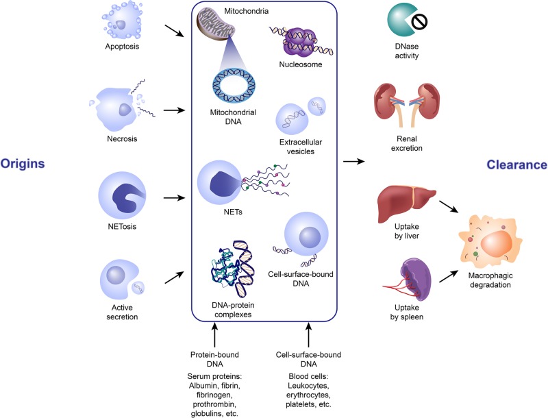
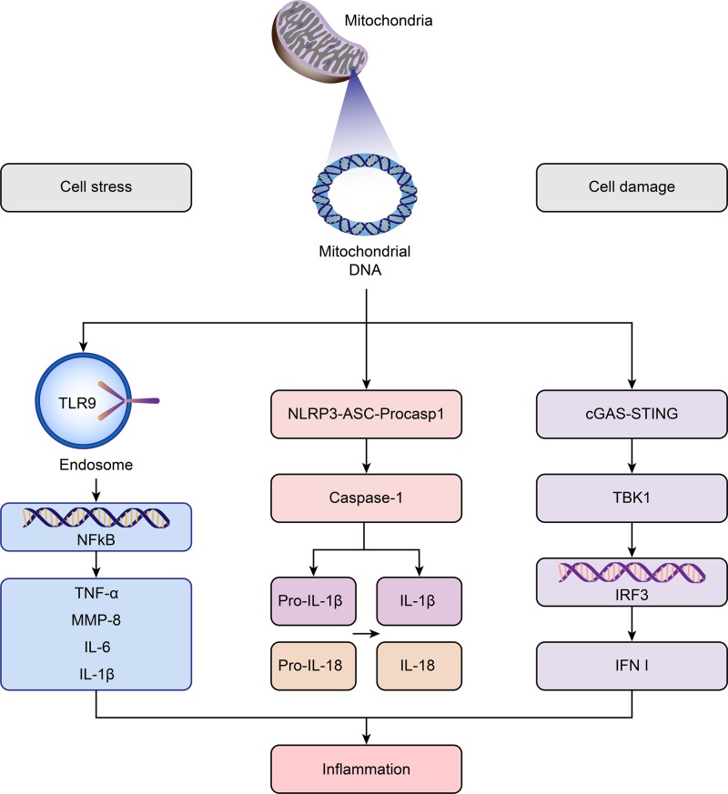
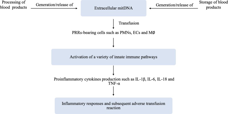
Blood transfusions are sometimes necessary after a high loss of blood due to injury or surgery. Some people need regular transfusions due to medical conditions such as haemophilia or cancer. Studies have suggested that extracellular DNA including mitochondrial DNA present in the extracellular milieu of transfused blood products has biological actions that are capable of activating the innate immune systems and potentially contribute to some adverse reactions in transfusion. From the present work, it becomes increasingly clear that extracellular DNA encompassed mitochondrial DNA is far from being biologically inert in blood products. It has been demonstrated to be present in eligible blood products and thus can be transfused to blood recipients. Although the presence of extracellular DNA in human plasma was initially detected in 1948, some aspects have not been fully elucidated. In this review, we summarize the potential origins, clearance mechanisms, relevant structures, and potential role of extracellular DNA in the innate immune responses and its relationship with individual adverse reactions in transfusion.

Keywords: extracellular DNA; horizontal gene transfer; innate immune response; mtDNA; transfusion; transfusion adverse reaction.


Conflict of interest statement

The authors declare that there are no competing interests associated with the manuscript.

Figures

Figure 1
Figure 1. The potential origins and clearance of extracellular DNA in blood products
ecDNA in blood exists in a number of forms, namely histone/DNA complexes or nucleosomes, extracellular vesicles packed DNA, cell-surface-bund DNA, DNA-protein complex, NETs and etc. It can be liberated from the blood cells via different mechanisms, most prominently apoptosis, necrosis, and active secretion, although other forms of cell death and clearance may contribute. Mitochondrial DNA can also be released by these mechanisms. Elimination of ecDNA could be achieved by DNase degradation, renal excretion into the urine or uptake by the liver and spleen followed by macrophagic degradation. Abbreviations: NETs, neutrophil extracellular traps.
Figure 2
Figure 2. Innate immune pathways activated by mitochondrial DNA
Upon cell stress or cell damage, mtDNA escaped from mitochondria engages in the activation of multiple innate immune pathways, including mitDNA–TLR9–NFκB axis, mitDNA–NLRP3–caspase1 pathway and mitDNA–cGAS–cGAMP–STING signaling, thereby igniting inflammation. Abbreviations: IFN, interferon; IRF, interferon regulatory factor; MMP, matrix metalloproteinase; NF-κB, nuclear transcription factor kappa B; NLRP3, Nod-like receptor pyrin domain containing 3; TBK1, TANK-binding kinase 1; TLR9, Toll-like receptor 9; TNF, tumor necrosis factor.
Figure 3
Figure 3. Working hypothesis of the involvement of extracellular mitDNA in the adverse transfusion reaction
Briefly, the released/generated extracellular mitDNA in blood products during processing and storage time, which can be sensed by PRR-bearing cells of recipients (i.e. PMNs, ECs and M∅), is capable of activating a variety of innate immune pathways. Subsequent production of cytokines (i.e. IL-1β, IL-6, IL-18, and TNF-α) together with other proinflammatory mediator substances, thereby inducing inflammatory responses leading to adverse transfusion reactions in recipients. Abbreviations: EC, endothelial cell; INF, interferon; M∅, macrophages; PMN, neutrophil; PRR, pattern recognition receptor; TNF, tumor necrosis factor.

Similar articles

Cited by

References

    1. Mandel P. and Métais P. (1948) Les acides nucléiques du plasma sanguin chez l'homme. C. R. Seances Soc. Biol. Fil. 142, 241–243 - PubMed
    1. Tamkovich S.N., Bryzgunova O.E., Rykova E.Y., Permyakova V.I., Vlassov V.V. and Laktionov P.P. (2005) Circulating nucleic acids in blood of healthy male and female donors. Clin. Chem. 51, 1317–1319 10.1373/clinchem.2004.045062 - DOI - PubMed
    1. Thierry A.R., El Messaoudi S., Gahan P.B., Anker P. and Stroun M. (2016) Origins, structures, and functions of circulating DNA in oncology. Cancer Metastasis Rev. 35, 347–376 10.1007/s10555-016-9629-x - DOI - PMC - PubMed
    1. Fernando M.R., Jiang C., Krzyzanowski G.D. and Ryan W.L. (2017) New evidence that a large proportion of human blood plasma cell-free DNA is localized in exosomes. PLoS ONE 12, e0183915 10.1371/journal.pone.0183915 - DOI - PMC - PubMed
    1. Kustanovich A., Schwartz R., Peretz T. and Grinshpun A. (2019) Life and death of circulating cell-free DNA. Cancer Biol. Ther. 20, 1057–1067 10.1080/15384047.2019.1598759 - DOI - PMC - PubMed

Publication types

MeSH terms