. 2020 Apr 1;318(4):F971-F978.
doi: 10.1152/ajprenal.00597.2019. Epub 2020 Mar 9.

Evaluation of cisplatin-induced injury in human kidney organoids

Affiliations

Evaluation of cisplatin-induced injury in human kidney organoids

Jenny L M Digby et al. Am J Physiol Renal Physiol. .

Abstract

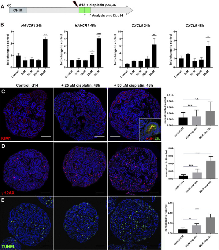
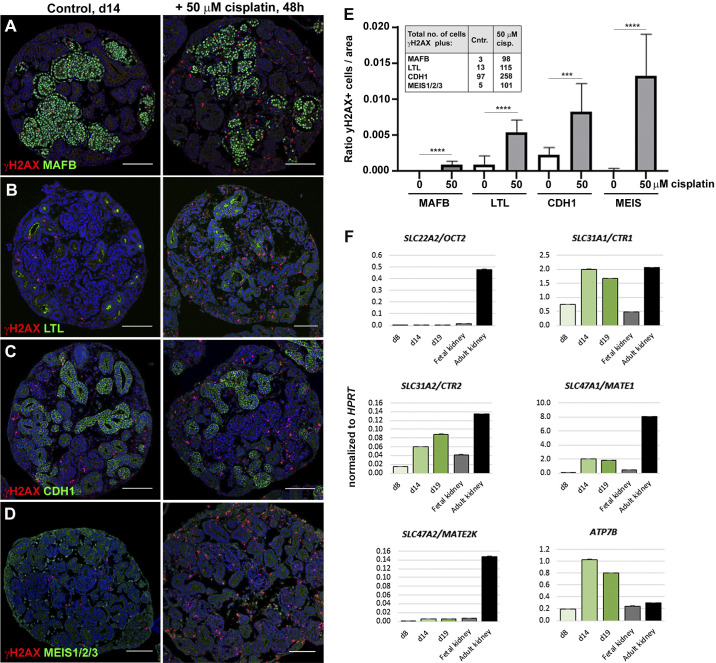
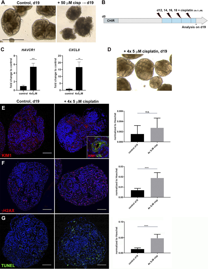
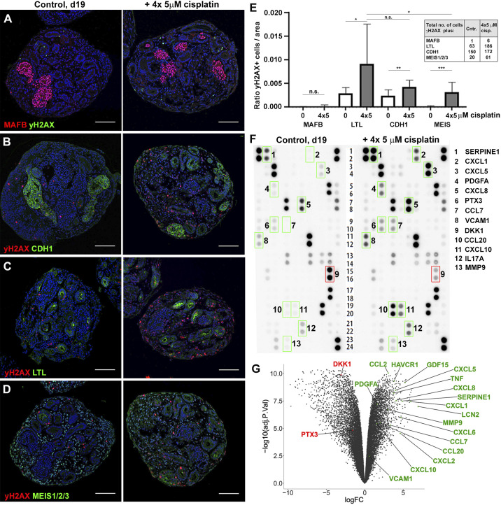
Acute kidney injury (AKI) remains a major global healthcare problem, and there is a need to develop human-based models to study AKI in vitro. Toward this goal, we have characterized induced pluripotent stem cell-derived human kidney organoids and their response to cisplatin, a chemotherapeutic drug that induces AKI and preferentially damages the proximal tubule. We found that a single treatment with 50 µM cisplatin induces hepatitis A virus cellular receptor 1 (HAVCR1) and C-X-C motif chemokine ligand 8 (CXCL8) expression, DNA damage (γH2AX), and cell death in the organoids but greatly impairs organoid viability. DNA damage was not specific to the proximal tubule but also affected the distal tubule and interstitial cell populations. This lack of specificity correlated with low expression of proximal tubule-specific SLC22A2/organic cation transporter 2 (OCT2) for cisplatin. To improve viability, we developed a repeated low-dose regimen of 4 × 5 µM cisplatin over 7 days and found this caused less toxicity while still inducing a robust injury response that included secretion of known AKI biomarkers and inflammatory cytokines. This work validates the use of human kidney organoids to model aspects of cisplatin-induced injury, with the potential to identify new AKI biomarkers and develop better therapies.

Keywords: acute kidney injury; acute kidney injury biomarker; cisplatin; cytokine; inflammation; kidney organoids; nephrotoxicity; proximal tubule; repeated low-dose regimen.


Conflict of interest statement

No conflicts of interest, financial or otherwise, are declared by the authors.

Figures

Fig. 1.
Fig. 1.
Cisplatin induces tubular injury, DNA damage, and cell death in kidney organoids. A: schematic of cisplatin treatment on kidney organoids. CHIR, chemical compound CHIR99021 used as in Ref. . B: quantitative PCR showing elevated expression of hepatitis A virus cellular receptor 1 (HAVCR1) and C-X-C motif chemokine ligand 8 (CXCL8) with increasing doses of cisplatin. C–E: immunohistochemistry staining on paraffin sections of day 14 organoids and quantification showing levels of tubular injury marker kidney injury molecule-1 [KIM1; colocalized to Lotus tetragonolobus lectin (LTL)+ proximal tubule; C, inset], DNA damage marker γH2AX, and cell death marker TUNEL increased with 25 and 50 µM cisplatin. d, day; ns, not significant. *P ≤ 0.05; **P ≤ 0.01; ****P ≤ 0.0001. Scale bars = 100 μm.
Fig. 2.
Fig. 2.
Cisplatin predominantly targets interstitial cells in kidney organoids. AD: immunohistochemistry staining for colocalization of DNA damage marker γH2AX with MAF BZIP transcription factor B (MAFB)+ podocytes, Lotus tetragonolobus lectin (LTL)+ proximal tubules, E-cadherin (CDH1)+ distal tubules, and MEIS1/2/3+ interstitial cells in control (Cntr) and 50 µM cisplatin-treated organoids. E: ratio of γH2AX+ cells per marker. Inset shows the total numbers of double-positive cells counted. F: quantitative PCR showing expression of cisplatin transporters in day 8 (d8), day 14 (d14), and day 19 (d19) kidney organoids and fetal and adult human kidneys. OCT2, organic cation transporter 2; CTR1 and CTR2, copper transporter 1 and 2, respectively; MATE1 and MATE2K, multidrug and toxin extrusion proteins 1 and 2, respectively; ATP7B, ATPase copper transporting-β; HPRT, hypoxanthine-guanine phosphoribosyltransferase 1. ***P ≤ 0.001; ****P ≤ 0.0001. Scale bars = 100 μm.
Fig. 3.
Fig. 3.
Repeated exposure to low-dose cisplatin reduces cell death and structural deterioration. A: bright-field imaging showing healthy organoids on day 19 (d19) and organoid deterioration after 50 µM cisplatin. B: schematic of repeated low-dose cisplatin treatment. C: quantitative PCR analysis of day 19 organoids showing hepatitis A virus cellular receptor 1 (HAVCR1) and C-X-C motif chemokine ligand 8 (CXCL8) expression increased upon 4 × 5 µM cisplatin. D: organoids treated with 4 × 5 µM cisplatin maintained tubular structures. EG: immunohistochemistry showing levels of kidney injury molecule-1 [KIM1; colocalized to Lotus tetragonolobus lectin (LTL)+ proximal tubule; E, inset], γH2AX, and TUNEL increased with 4 × 5 µM cisplatin. ns, not significant. **P ≤ 0.01; ***P ≤ 0.001; ****P ≤ 0.0001. Scale bars = 400 μm in A and 100 μm in EG.
Fig. 4.
Fig. 4.
Organoids secrete acute kidney injury biomarkers and cytokines in response to cisplatin. AD: immunohistochemistry on day 19 (d19) control (Cntr) and 4 × 5 µM cisplatin-treated organoids for colocalization of DNA damage (γH2AX) with kidney tissues [MAF BZIP transcription factor B (MAFB) + podocytes, Lotus tetragonolobus lectin (LTL)+ proximal tubules, E-cadherin (CDH1)+ distal tubules, and MEIS1/2/3+ interstitial cells]. E: quantification of γH2AX colocalization with kidney tissues. Inset shows total numbers of double-positive cells counted. F: cytokine array analysis using culture media collected from control and 4 × 5 µM cisplatin-treated organoids. Factors with higher (green boxes) or lower (red box) secretion in cisplatin-treated versus control organoids are shown. G: volcano plot showing the result of RNA sequencing profiling of control and 4 × 5 µM cisplatin-treated organoids. The genes encoding the differentially secreted cytokines and a selection of acute kidney injury biomarkers that are differentially expressed upon cisplatin treatment are highlighted. ns, not significant. See text for abbreviations. *P ≤ 0.05; **P ≤ 0.01; ***P ≤ 0.001. Scale bars = 100 μm.

References

    1. Black LM, Lever JM, Traylor AM, Chen B, Yang Z, Esman SK, Jiang Y, Cutter GR, Boddu R, George JF, Agarwal A. Divergent effects of AKI to CKD models on inflammation and fibrosis. Am J Physiol Renal Physiol 315: F1107–F1118, 2018. doi: 10.1152/ajprenal.00179.2018. - DOI - PMC - PubMed
    1. Ciarimboli G, Ludwig T, Lang D, Pavenstädt H, Koepsell H, Piechota H-J, Haier J, Jaehde U, Zisowsky J, Schlatter E. Cisplatin nephrotoxicity is critically mediated via the human organic cation transporter 2. Am J Pathol 167: 1477–1484, 2005. doi: 10.1016/S0002-9440(10)61234-5. - DOI - PMC - PubMed
    1. Cortvrindt C, Speeckaert R, Moerman A, Delanghe JR, Speeckaert MM. The role of interleukin-17A in the pathogenesis of kidney diseases. Pathology 49: 247–258, 2017. doi: 10.1016/j.pathol.2017.01.003. - DOI - PubMed
    1. Dobin A, Davis CA, Schlesinger F, Drenkow J, Zaleski C, Jha S, Batut P, Chaisson M, Gingeras TR. STAR: ultrafast universal RNA-seq aligner. Bioinformatics 29: 15–21, 2013. doi: 10.1093/bioinformatics/bts635. - DOI - PMC - PubMed
    1. Freedman BS, Brooks CR, Lam AQ, Fu H, Morizane R, Agrawal V, Saad AF, Li MK, Hughes MR, Werff RV, Peters DT, Lu J, Baccei A, Siedlecki AM, Valerius MT, Musunuru K, McNagny KM, Steinman TI, Zhou J, Lerou PH, Bonventre JV. Modelling kidney disease with CRISPR-mutant kidney organoids derived from human pluripotent epiblast spheroids. Nat Commun 6: 8715, 2015. doi: 10.1038/ncomms9715. - DOI - PMC - PubMed

Publication types

MeSH terms