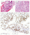
Composite Chronic Lymphocytic Leukemia/Small Lymphocytic Lymphoma and Mantle Cell Lymphoma; Small Cell Variant: A Real Diagnostic Challenge. Case Presentation and Review of Literature
- PMID: 32150530
- PMCID: PMC7083592
- DOI: 10.12659/AJCR.921131
Composite Chronic Lymphocytic Leukemia/Small Lymphocytic Lymphoma and Mantle Cell Lymphoma; Small Cell Variant: A Real Diagnostic Challenge. Case Presentation and Review of Literature
Abstract
BACKGROUND Chronic lymphocytic leukemia/small lymphocytic lymphoma (CLL/SLL) and mantle cell lymphoma (MCL) both have a common origin arising from mature CD5+ B-lymphocytes. Their distinction is crucial since MCL is a considerably more aggressive disease. Composite lymphoma consisting of CLL/SLL and MCL has been rarely reported. This type of composite lymphoma may be under-diagnosed as the 2 neoplasms have many features in common, both morphologically and immunophenotypically. CASE REPORT We report the case of a 57-year-old male patient who presented with a 4-month history of recurrent abdominal pain and distention with hepatosplenomegaly. Peripheral blood showed a high leukocytes count (46.7×10³/uL) with marked lymphocytosis of 35.0×10³/uL, mostly small mature-looking, with some showing nuclear irregularities, with approximately 3% prolymphocytes. Immunophenotyping by flow cytometry and immunohistochemistry revealed 2 immunophenotypically distinct abnormal CD5+monotypic B-cell populations. Fluorescence in situ hybridization (FISH) on peripheral blood demonstrated IGH/CCND1 rearrangement consistent with t(11;14) in 65% of cells analyzed. Accordingly, based on compilation of findings from morphology, flow cytometry, immunohistochemistry, and FISH, A diagnosis of composite lymphoma consisting of MCL; small cell variant and CLL/SLL was concluded. CONCLUSIONS We describe a case of composite lymphoma of MCL (small cell variant) and CLL/SLL that emphasizes the crucial role of the multiparametric approach, including vigilant cyto-histopathologic examination, immunophenotyping by flow cytometry and immunohistochemistry, as well as genetic testing, to achieve the correct diagnosis.
Conflict of interest statement
None.
Figures


Similar articles
-
Composite mantle cell lymphoma and chronic lymphocytic leukemia/small lymphocytic lymphoma: a clinicopathologic and molecular study.Hum Pathol. 2013 Jan;44(1):110-21. doi: 10.1016/j.humpath.2012.04.022. Epub 2012 Aug 31. Hum Pathol. 2013. PMID: 22944294 Free PMC article. Review.
-
Mantle cell lymphoma with chronic lymphocytic leukemia-like features: a diagnostic mimic and pitfall.Hum Pathol. 2022 Jan;119:59-68. doi: 10.1016/j.humpath.2021.11.001. Epub 2021 Nov 9. Hum Pathol. 2022. PMID: 34767860
-
Composite mantle cell lymphoma and chronic lymphocytic leukemia/small lymphocytic lymphoma.Cytometry B Clin Cytom. 2018 Jan;94(1):148-150. doi: 10.1002/cyto.b.21512. Epub 2017 Feb 24. Cytometry B Clin Cytom. 2018. PMID: 28109040
-
Diagnosis and prognosis of B-cell chronic lymphocytic leukemia/small lymphocytic lymphoma (B-CLL/SLL) and mantle cell lymphoma (MCL).J Egypt Natl Canc Inst. 2005 Dec;17(4):279-90. J Egypt Natl Canc Inst. 2005. PMID: 17102815
-
Differential diagnosis of chronic lymphocytic leukemia/small lymphocytic lymphoma and other indolent lymphomas, including mantle cell lymphoma.J Clin Exp Hematop. 2020 Dec 15;60(4):124-129. doi: 10.3960/jslrt.19041. Epub 2020 Apr 3. J Clin Exp Hematop. 2020. PMID: 32249238 Free PMC article. Review.
Cited by
-
Hodgkin Lymphoma and Hairy Cell Leukemia Arising from Chronic Lymphocytic Leukemia: Case Reports and Literature Review.J Clin Med. 2022 Aug 10;11(16):4674. doi: 10.3390/jcm11164674. J Clin Med. 2022. PMID: 36012912 Free PMC article.
-
Composite mantle cell and Burkitt lymphoma: a rare case report.Porto Biomed J. 2024 Jun 20;9(3):257. doi: 10.1097/j.pbj.0000000000000257. eCollection 2024 May-Jun. Porto Biomed J. 2024. PMID: 38903392 Free PMC article. No abstract available.
-
Concurrent chronic lymphocytic leukemia/small lymphocytic lymphoma and hairy cell leukemia: clinical, pathologic and molecular features.Leuk Lymphoma. 2020 Dec;61(13):3177-3187. doi: 10.1080/10428194.2020.1797007. Epub 2020 Aug 5. Leuk Lymphoma. 2020. PMID: 32755330 Free PMC article.
-
Biclonal Low-Grade B Cell Lymphoma as Detected by Multiparametric Flow Cytometry in Blood and Marrow.Indian J Hematol Blood Transfus. 2023 Oct;39(4):691-698. doi: 10.1007/s12288-022-01625-y. Epub 2023 Jan 2. Indian J Hematol Blood Transfus. 2023. PMID: 37786829 Free PMC article.
-
Hemophagocytic lymphohistiocytosis secondary to composite lymphoma: Two case reports.World J Clin Cases. 2021 Oct 26;9(30):9159-9167. doi: 10.12998/wjcc.v9.i30.9159. World J Clin Cases. 2021. PMID: 34786400 Free PMC article.
References
-
- Swerdlow SH, Campo E, Seto M, Mller-Hemelink HK. Mantle cell lymphoma in WHO classification of tumors of haemopoietic and lymphoid tissue. Revised 4th Edition. Lyon; 2017.
-
- Babbage G, Garand R, Robillard N, et al. Mantle cell lymphoma with t(11;14) and unmutated or mutated VH genes expresses AID and undergoes isotype switch events. Blood. 2004;103:2795–98. - PubMed
-
- Igawa T, Omote R, Sato H, et al. A possible new morphological variant of mantle cell lymphoma with plasma-cell type Castleman disease-like features. Pathol Res Pract. 2017;213(11):1378–83. - PubMed
-
- Campo E, Ghia P, Montserrat E, et al. Chronic lymphocytic leukaemia/small lymphocytic lymphoma in WHO classification of tumors of haemopoietic and lymphoid tissue. Revised 4th Edition. Lyon; 2017.
-
- Siegel RL, Miller KD, Jemal A. Cancer statistics. Cancer J Clin. 2018;68(1):7. - PubMed
Publication types
MeSH terms
Substances
LinkOut - more resources
Full Text Sources
Research Materials

