. 2020 Mar 9;16(3):e1008659.
doi: 10.1371/journal.pgen.1008659. eCollection 2020 Mar.

A putative silencer variant in a spontaneous canine model of retinitis pigmentosa

Collaborators, Affiliations

A putative silencer variant in a spontaneous canine model of retinitis pigmentosa

Maria Kaukonen et al. PLoS Genet. .

Abstract

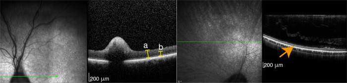
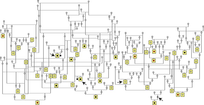
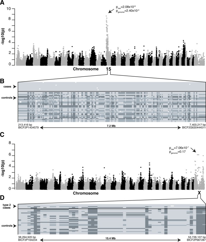
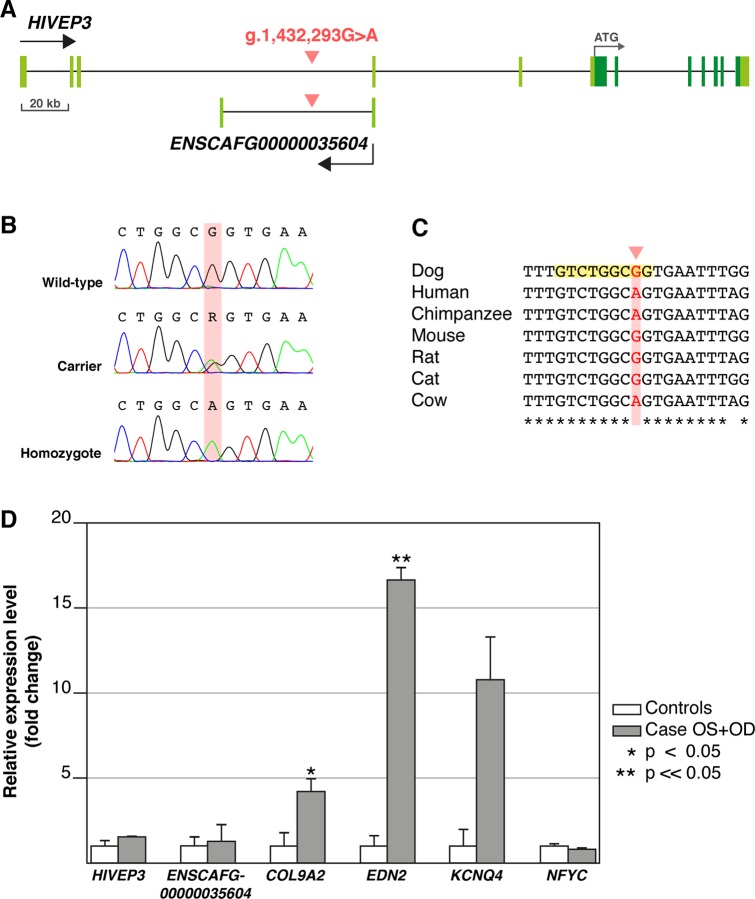
Retinitis pigmentosa (RP) is the leading cause of blindness with nearly two million people affected worldwide. Many genes have been implicated in RP, yet in 30-80% of the RP patients the genetic cause remains unknown. A similar phenotype, progressive retinal atrophy (PRA), affects many dog breeds including the Miniature Schnauzer. We performed clinical, genetic and functional experiments to identify the genetic cause of PRA in the breed. The age of onset and pattern of disease progression suggested that at least two forms of PRA, types 1 and 2 respectively, affect the breed, which was confirmed by genome-wide association study that implicated two distinct genomic loci in chromosomes 15 and X, respectively. Whole-genome sequencing revealed a fully segregating recessive regulatory variant in type 1 PRA. The associated variant has a very recent origin based on haplotype analysis and lies within a regulatory site with the predicted binding site of HAND1::TCF3 transcription factor complex. Luciferase assays suggested that mutated regulatory sequence increases expression. Case-control retinal expression comparison of six best HAND1::TCF3 target genes were analyzed with quantitative reverse-transcriptase PCR assay and indicated overexpression of EDN2 and COL9A2 in the affected retina. Defects in both EDN2 and COL9A2 have been previously associated with retinal degeneration. In summary, our study describes two genetically different forms of PRA and identifies a fully penetrant variant in type 1 form with a possible regulatory effect. This would be among the first reports of a regulatory variant in retinal degeneration in any species, and establishes a new spontaneous dog model to improve our understanding of retinal biology and gene regulation while the affected breed will benefit from a reliable genetic testing.


Conflict of interest statement

We have read the journal’s policy and the authors of this manuscript have the following competing interests: HL is a paid consultant to Genoscoper Laboratories Ltd, which will provide a genetic test for the type 1 PRA in MSs.

Figures

Fig 1
Fig 1. PRA confirmation by optical coherence tomography (OCT).
OCT assessment of a healthy control (left hand side) and a four-year-old MS with severe type 1 PRA findings (right hand side). Whole retinal (a) and photoreceptor inner and outer segment and outer nuclear layer thickness (b) measurement showed total loss of the photoreceptor cell layers in the affected dog (orange arrow).
Fig 2
Fig 2. Pedigree of the study cohort.
Pedigree analysis suggests a recessive mode of inheritance as affected dogs were born to unaffected parents. Squares indicate males and circles females. Individuals marked with black are type 1 cases, while type 2 cases are marked with red. Dogs with yellow background were genotyped and included in GWA study. Black arrows mark the whole-genome sequenced dogs.
Fig 3
Fig 3. Mapping PRA loci in MSs.
(A) Genome-wide comparison of allele frequencies in type 1 cases (n = 10) and controls (n = 33) indicated a locus on the CFA15 (praw = 2.08x10⁻⁹, pgenome = 2.40x10⁻⁴). (B) A shared homozygous haplotype block was seen in all the type 1 cases and one control and absent in 32/33 controls. Each row represents a single animal while genotypes at each SNP (columns) are marked with light (homozygous), dark (opposite homozygous) or intermediate (heterozygotes) grey. The critical region of 7.2 Mb spans from 213,416 bp to 7,403,217 bp and upper and lower limits of it were determined by appearance of heterozygous SNPs in the case dogs. (C) GWAS in type 2 cases (n = 6) and controls (n = 33) suggests a locus on CFAX (praw = 7.06x10⁻⁷, pgenome = 0.17). (D) A shared homo- or hemizygous haplotype block of 15.4 Mb (spanning nucleotides 38,294,920–53,726,107 bp) is present in 3 out of 6 type 2 cases and absent in the controls and the rest of the type 2 cases.
Fig 4
Fig 4. Schematic representation of the variant (g.1,432,293G>A) site and RT-qPCR results of HAND1::TCF3 target genes.
(A) The HIVEP3 and ENSCAFG00000035604 gene structures with coding regions indicated in dark and non-coding in light green. The intronic variant (red) is located kilobases away of the exon-intron boundaries in both genes. (B) Sanger chromatograms showing the DNA sequence in wild-type, carrier and homozygous dogs with the mutated nucleotide marked with red background. (C) Alignment of different mammalian species showed that the sequence surrounding the variant site is highly conserved, but the variant nucleotide itself (highlighted in red) is not. JASPAR motif scanning indicated that TFBS of HAND1::TCF3 overlaps exactly the variant site (highlighted in yellow). (D) RT-qPCR of retinal samples from a type 1 affected MS and four control retinal samples wild-type for the variant and without the studied phenotype showed case-specific over-expression of EDN2 and COL9A2. The comparative ΔΔCT method was used to determine relative expression [24] and error bars are determined as standard deviation of the mean ΔCT.
Fig 5
Fig 5. Reporter assay indicates the variant site might be a silencer.
Dual luciferase assay with MDCK cells transfected with wild-type (WT), mutant (MT) and empty vectors indicated robust reduction in relative luciferase activity when comparing the wild-type constructs to empty vector, supporting the putative silencer activity (p < 0.05). Relative luciferase activity was significantly increased in mutant versus wild-type constructs (p < 0.05), indicating the type 1 PRA associated variant disrupts the putative silencer activity, but might not prevent it completely.

Comment in

  • Formal commentary.
    Aguirre GD, Lohi H, Kaukonen M, Murgiano L. Aguirre GD, et al. PLoS Genet. 2020 Nov 5;16(11):e1009059. doi: 10.1371/journal.pgen.1009059. eCollection 2020 Nov. PLoS Genet. 2020. PMID: 33151924 Free PMC article. No abstract available.

Similar articles

Cited by

References

    1. Narayan DS, Wood JP, Chidlow G, Casson RJ. A review of the mechanisms of cone degeneration in retinitis pigmentosa. Acta Ophthalmol 2016;94(8):748–754. 10.1111/aos.13141 - DOI - PubMed
    1. Daiger S, Rossiter B, Greenberg J, Christoffels A, Hide W. Data services and software for identifying genes and mutations causing retinal degeneration. Invest Ophthalmol Vis Sci 1998;39:S295.
    1. Daiger S, Sullivan L, Bowne S. Genes and mutations causing retinitis pigmentosa. Clin Genet 2013;84(2):132–141. 10.1111/cge.12203 - DOI - PMC - PubMed
    1. Bassuk AG, Zheng A, Li Y, Tsang SH, Mahajan VB. Precision Medicine: Genetic Repair of Retinitis Pigmentosa in Patient-Derived Stem Cells. Sci Rep 2016. January 27;6:19969 10.1038/srep19969 - DOI - PMC - PubMed
    1. Lindblad-Toh K, Wade CM, Mikkelsen TS, Karlsson EK, Jaffe DB, Kamal M, et al. Genome sequence, comparative analysis and haplotype structure of the domestic dog. Nature 2005;438(7069):803–819. 10.1038/nature04338 - DOI - PubMed

Publication types

MeSH terms