. 2020 Mar 9;16(3):e1008392.
doi: 10.1371/journal.ppat.1008392. eCollection 2020 Mar.

Structure of mouse coronavirus spike protein complexed with receptor reveals mechanism for viral entry

Affiliations

Structure of mouse coronavirus spike protein complexed with receptor reveals mechanism for viral entry

Jian Shang et al. PLoS Pathog. .

Abstract

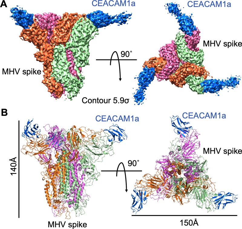
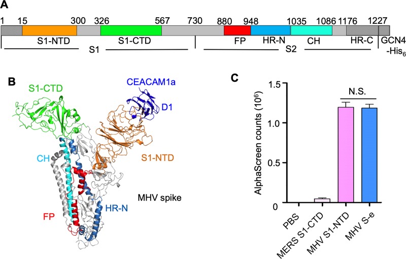
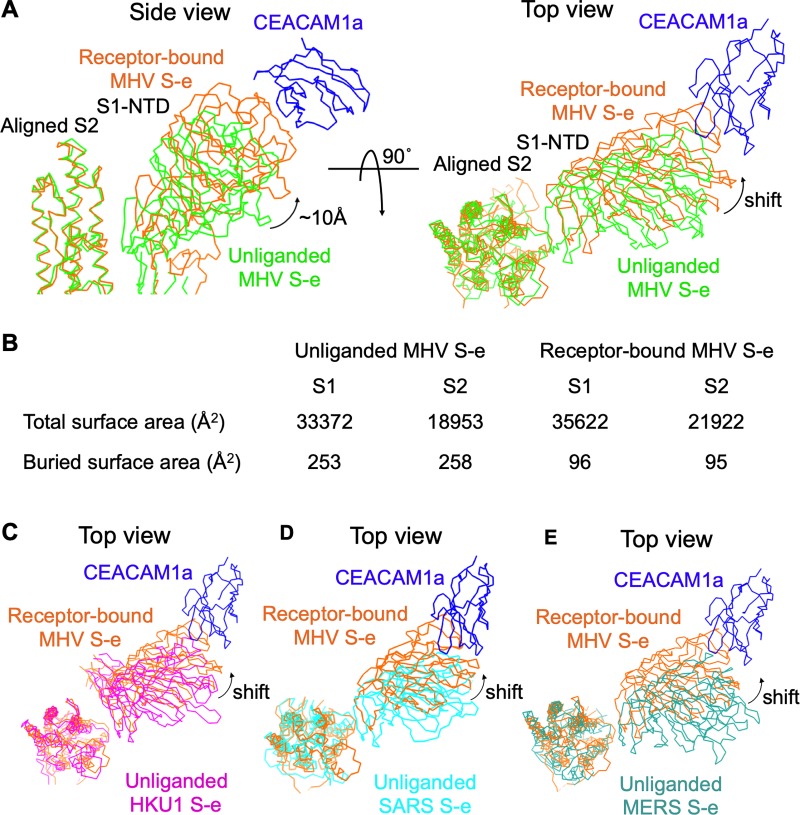
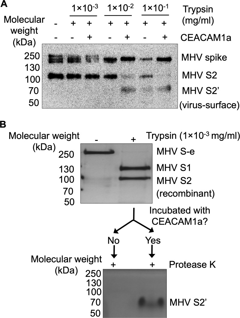
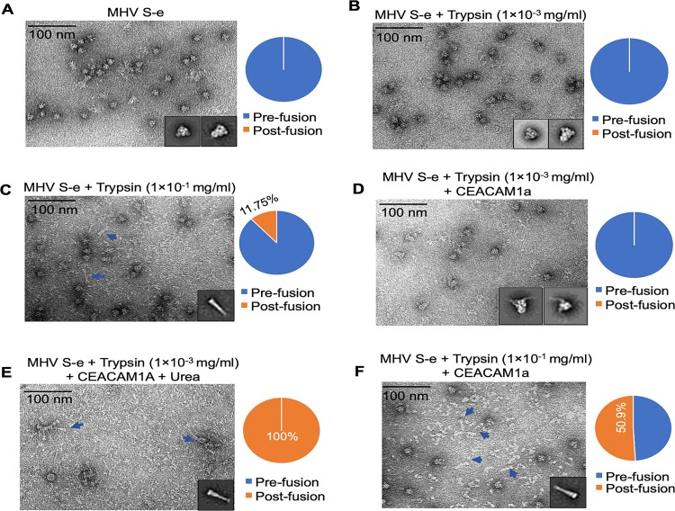
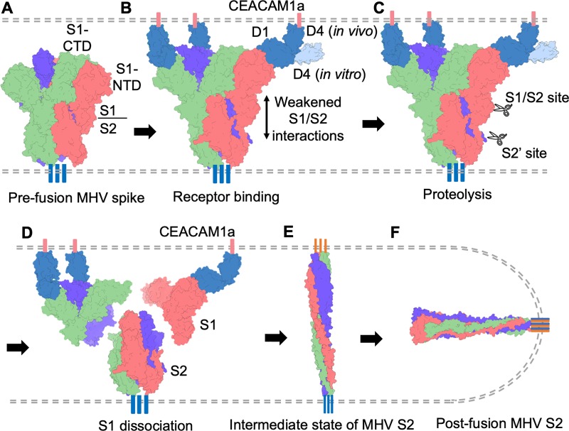
Coronaviruses recognize a variety of receptors using different domains of their envelope-anchored spike protein. How these diverse receptor recognition patterns affect viral entry is unknown. Mouse hepatitis coronavirus (MHV) is the only known coronavirus that uses the N-terminal domain (NTD) of its spike to recognize a protein receptor, CEACAM1a. Here we determined the cryo-EM structure of MHV spike complexed with mouse CEACAM1a. The trimeric spike contains three receptor-binding S1 heads sitting on top of a trimeric membrane-fusion S2 stalk. Three receptor molecules bind to the sides of the spike trimer, where three NTDs are located. Receptor binding induces structural changes in the spike, weakening the interactions between S1 and S2. Using protease sensitivity and negative-stain EM analyses, we further showed that after protease treatment of the spike, receptor binding facilitated the dissociation of S1 from S2, allowing S2 to transition from pre-fusion to post-fusion conformation. Together these results reveal a new role of receptor binding in MHV entry: in addition to its well-characterized role in viral attachment to host cells, receptor binding also induces the conformational change of the spike and hence the fusion of viral and host membranes. Our study provides new mechanistic insight into coronavirus entry and highlights the diverse entry mechanisms used by different viruses.


Conflict of interest statement

The authors have declared that no competing interests exist.

Figures

Fig 1
Fig 1. Overall structure of MHV spike protein/CEACAM1a complex.
(A) Cryo-EM density map of MHV spike ectodomain/CEACAM1a complex. Left: side view. Right: top view. The trimeric MHV spike ectodomain (S-e) is in the pre-fusion state. Each monomeric subunit of MHV S-e is colored differently and CEACAM1A is colored in blue. (B) Atomic structure of MHV S-e/CEACAM1a complex. The molecules and subunits are colored in the same way as in panel (A). The views are also the same as in panel (A). The D4 domain of CEACAM1a had weak densities and hence its atomic model was not built.
Fig 2
Fig 2. Detailed structure of MHV spike protein/CEACAM1a complex.
(A) Schematic drawing of MHV S-e. S1: receptor-binding subunit. S2: membrane-fusion subunit. GCN4-His6: GCN4 trimerization tag followed by His6 tag. S1-NTD: N-terminal domain of S1. S1-CTD: C-terminal domain of S1. CH: central helix. FP: fusion peptide. HR-N and HR-C: heptad repeats N and C, respectively. (B) Structure of a monomeric subunit of MHV S-e/CEACAM1a complex. The structural elements of MHV S-e are colored in the same way as in panel (A). CEACAM1a is colored in blue. (C) Binding interactions between recombinant CEACAM1a (with a C-terminal Fc tag) and recombinant MHV S1-NTD or recombinant MHV S-e (with a C-terminal His6 tag) were measured using AlphaScreen assay. PBS and MERS-CoV S1-CTD, neither of which binds CEACAM1a, served as negative controls for MHV S-e and MHV S1-NTD. The error bars indicate standard deviation (SD) (n = 5). N.S.: statistically not significant (P > 0.05 in two tailed t-test).
Fig 3
Fig 3. CEACAM1a-induced structural change of MHV spike.
(A) Comparison of chain traces of S1-NTD in receptor-bound MHV S-e (colored in orange) and that in unliganded MHV S-e (colored in green), with the S2 subunits from the two S-e molecules aligned together. (B) Comparison of buried surface areas of S1 and S2 in receptor-bound MHV S-e trimer and unliganded MHV S-e trimer. Here the S1 and S2 are defined as regions before and after residue 730 (Fig 2A), respectively. (C-E) Same as in panel (A), except that unliganded MHV S-e is replaced by unliganded HKU1 S-e (PDB ID: 5I08; colored in magenta; panel C), unliganded SARS-CoV S-e (PDB ID: 5X5F; colored in cyan; panel D), or unliganded MERS-CoV S-e (PDB ID: 5X8; colored in dark green; panel E).
Fig 4
Fig 4. Receptor-facilitated proteolysis of MHV spike.
(A) Western blot analysis of virus-surface MHV spike that had been cleaved by trypsin in the presence or absence of CEACAM1a. Different concentrations of trypsin were used. Here only protein fragments containing the C-terminal C9 tag (i.e., MHV spike, S2 and S2’, but not S1) could be detected since an antibody targeting the C-terminal C9 tag of MHV spike was used for the Western blot analysis. The result showed that receptor binding enhanced the protease sensitivity of MHV spike and produced more cleaved fragments (particularly S2’). (B) Silver staining analyses of recombinant MHV S-e that had been subjected to a double proteolysis assay. Specifically, recombinant MHV S-e molecules were first treated with low concentration of trypsin. Then half of the trypsin-cleaved products were incubated with CEACAM1a, while the other half were not. Subsequently both halves were treated with protease K. Here all protein fragments (i.e., MHV S-e, S1, S2 and S2’) could be detected as silver staining was used for the detection. The result showed that receptor treatment of the trypsin-cleaved MHV S-e led to a protease K-resistant S2’ fragment, suggesting that CEACAM1a binding facilitated the already cleaved MHV S-e to transition from pre-fusion to post-fusion conformation. See text for more discussion.
Fig 5
Fig 5. Negative-stain EM image of MHV spike treated with protease in the presence or absence of CEACAM1a.
(A) MHV S-e without any protease treatment. All of the S-e molecules were in the pre-fusion state. (B) MHV S-e treated with low concentration of trypsin. All of the S-e molecules were in the pre-fusion state. (C) MHV S-e treated with high concentration of trypsin. 11.75% of the S-e molecules were in the post-fusion conformation (featured by the rod-like structure). (D) MHV S-e treated with low concentration of trypsin and incubated with CECAAM1a. All of the S-e molecules were in the pre-fusion state. (E) MHV S-e treated with low concentration of trypsin and incubated with urea. All of the S-e molecules were in the post-fusion state. (F) MHV S-e treated with high concentration of trypsin and incubated with CEACAM1a. 50.9% of the S-e molecules were in the post-fusion conformation. 2D averages of the S-e particles were shown as insets of each panel.
Fig 6
Fig 6. Proposed molecular mechanism of MHV entry.
(A) Virus-surface MHV spike in the pre-fusion state. Each monomeric subunit of MHV spike trimer is colored differently. (B) Receptor binding by MHV spike. Host cell-surface CEACAM1a is colored in blue. Receptor binding triggers conformational changes in MHV spike, weakening the S1/S2 interactions. Although in vitro the receptor binds to MHV spike in an angle perpendicular to the spike, in vivo the receptor would need to bend in order to approach the receptor-binding sites in MHV spike. (C) Receptor-bound MHV spike is cleaved by proteases at two sites: S1/S2 site and S2' site. (D) Receptor facilitates S1 to dissociate from S2 through receptor-induced conformational changes in the spike, tension generated by potential bending of the receptor, and receptor-facilitated proteolysis of the spike. (E) Hypothetical intermediate state of MHV spike as proposed by many previous studies. (F) MHV spike transitions to the post-fusion state, leading to membrane fusion.

Similar articles

Cited by

References

    1. Li F. Receptor recognition mechanisms of coronaviruses: a decade of structural studies. J Virol. 2015;89(4):1954–64. Epub 2014/11/28. 10.1128/JVI.02615-14 - DOI - PMC - PubMed
    1. Li F. Structure, Function, and Evolution of Coronavirus Spike Proteins. Annual review of virology. 2016;3(1):237–61. Epub 2016/09/01. 10.1146/annurev-virology-110615-042301 . - DOI - PMC - PubMed
    1. Perlman S, Netland J. Coronaviruses post-SARS: update on replication and pathogenesis. Nature Reviews Microbiology. 2009;7(6):439–50. 10.1038/nrmicro2147 WOS:000266451100012. - DOI - PMC - PubMed
    1. Spaan W, Cavanagh D, Horzinek MC. Coronaviruses: structure and genome expression. J Gen Virol. 1988;69 (Pt 12):2939–52. Epub 1988/12/01. 10.1099/0022-1317-69-12-2939 . - DOI - PubMed
    1. Gonzaalez JM, Gomez-Puertas P, Cavanagh D, Gorbalenya AE, Enjuanes L. A comparative sequence analysis to revise the current taxonomy of the family Coronaviridae. Archives of Virology. 2003;148(11):2207–35. 10.1007/s00705-003-0162-1 WOS:000186399900009. - DOI - PMC - PubMed

Publication types

MeSH terms

Substances