. 2020 Mar 9;15(3):e0229769.
doi: 10.1371/journal.pone.0229769. eCollection 2020.

Neuroprotective effect of menaquinone-4 (MK-4) on transient global cerebral ischemia/reperfusion injury in rat

Affiliations

Neuroprotective effect of menaquinone-4 (MK-4) on transient global cerebral ischemia/reperfusion injury in rat

Bahram Farhadi Moghadam et al. PLoS One. .

Abstract

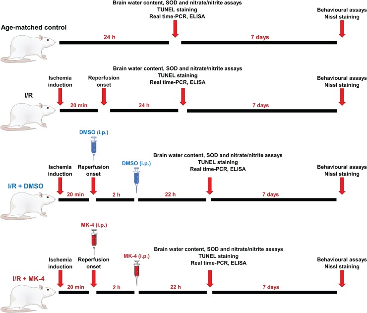
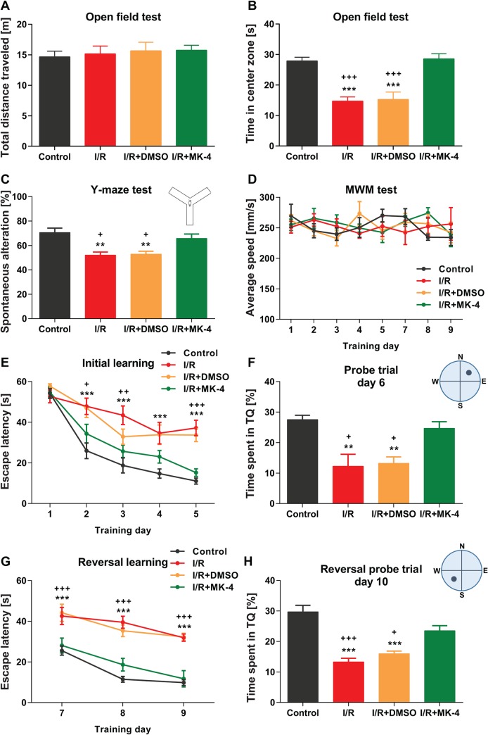
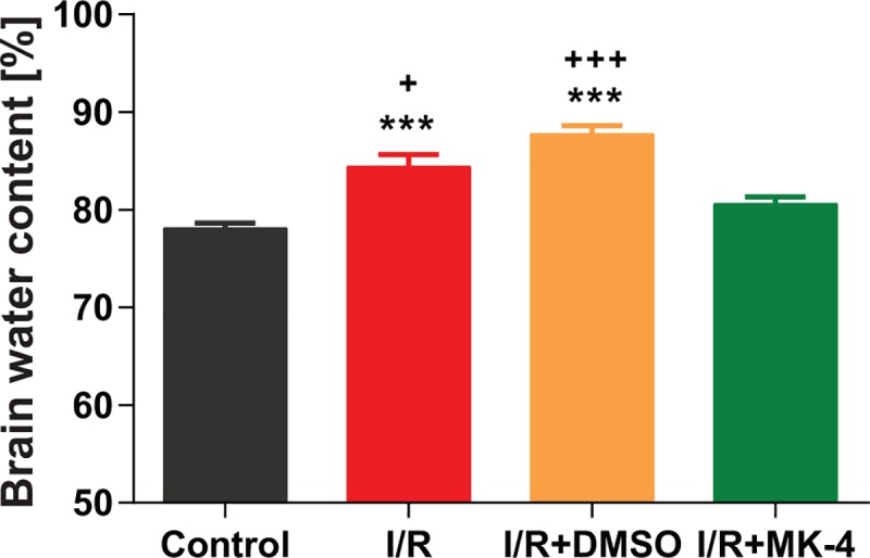
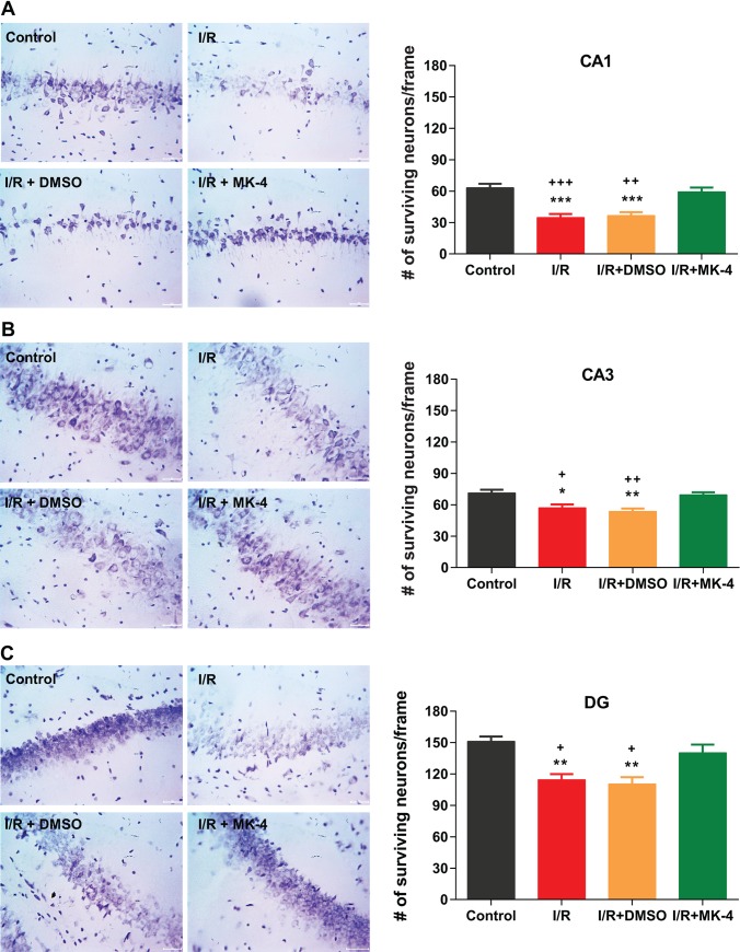
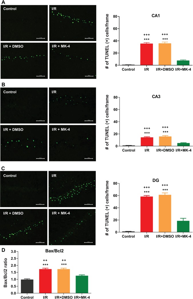
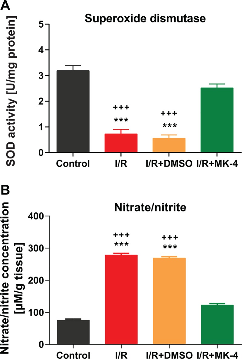
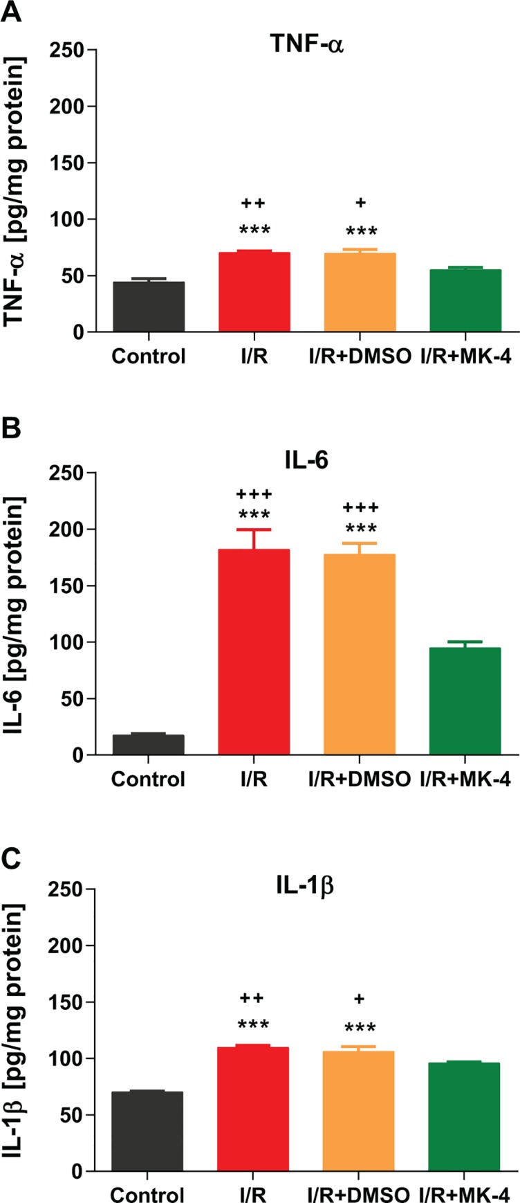
Cerebral ischemia/reperfusion (I/R) injury causes cognitive deficits, excitotoxicity, neuroinflammation, oxidative stress and brain edema. Vitamin K2 (Menaquinone 4, MK-4) as a potent antioxidant can be a good candidate to ameliorate I/R consequences. This study focused on the neuroprotective effects of MK-4 for cerebral I/R insult in rat's hippocampus. The rat model of cerebral I/R was generated by transient bilateral common carotid artery occlusion for 20 min. Rats were divided into control, I/R, I/R+DMSO (solvent (1% v/v)) and I/R+MK-4 treated (400 mg/kg, i.p.) groups. Twenty-four hours after I/R injury induction, total brain water content, superoxide dismutase (SOD) activity, nitrate/nitrite concentration and neuronal density were evaluated. In addition to quantify the apoptosis processes, TUNEL staining, as well as expression level of Bax and Bcl2, were assessed. To evaluate astrogliosis and induced neurotoxicity by I/R GFAP and GLT-1 mRNA expression level were quantified. Furthermore, pro-inflammatory cytokines including IL-1β, IL-6 and TNF-α were measured. Seven days post I/R, behavioral analysis to quantify cognitive function, as well as Nissl staining for surviving neuronal evaluation, were conducted. The findings indicated that administration of MK-4 following I/R injury improved anxiety-like behavior, short term and spatial learning and memory impairment induced by I/R. Also, MK-4 was able to diminish the increased total brain water content, apoptotic cell density, Bax/ Bcl2 ratio and GFAP mRNA expression following I/R. In addition, the high level of nitrate/nitrite, IL-6, IL-1β and TNF-α induced by I/R was reduced after MK-4 administration. However, MK-4 promotes the level of SOD activity and GLT-1 mRNA expression in I/R rat model. The findings demonstrated that MK-4 can rescue transient global cerebral I/R consequences via its anti-inflammatory and anti-oxidative stress features. MK-4 administration ameliorates neuroinflammation, neurotoxicity and neuronal cell death processes and leads to neuroprotection.


Conflict of interest statement

The authors have declared that no competing interests exist.

Figures

Fig 1
Fig 1. A schematic experimental timeline (N = 5–6 for each group).
Fig 2
Fig 2. Positive effect of 400 mg/kg MK-4 administration on mortality rate in I/R rat model.
Although administration of 100 mg/kg MK-4 following I/R showed a similar mortality rate compared to I/R group. The dose of 400 mg/kg MK-4 in two injections (200 mg/kg i.p. immediately and 200 mg/kg i.p. 2h after reperfusion) led to reduced mortality rate compared to I/R group.
Fig 3
Fig 3. The positive effects of MK-4 administration on behavioral deficits induced by I/R injury.
(A) In the open field test, total distance traveled was not changed significantly following I/R injury induction. (B) Time spent in the center zone of open field arena was reduced post I/R, however, i.p. administration of 400 mg/kg MK-4 (200 mg/kg immediately and 2 h after I/R injury) led to compensate the phenotype. (C) Spontaneous alternation behavior in the Y maze test was reduced in rat I/R model, but it backed to the control level following MK-4 administration in I/R animals. (D) In the Morris water maze test, swimming speed was not altered significantly between groups. (E-H) Escape latency during the initial (E-F) and reversal phases (G-H) of Morris water maze was higher in I/R animals. In addition, I/R rats spent less time in target quadrants (TQ) in both reference memory tests compared to control. Following MK-4 administration in I/R animals, the phenotypes disappeared and the results were comparable to control group. Data are presented as mean±SEM. ** p <0.01 and *** p < 0.001 compared to control, + p < 0.05, ++ p < 0.001 and +++ p < 0.001 compared to I/R+MK4 group (n = 6).
Fig 4
Fig 4. Reduction of brain water content following MK-4 application in I/R rat model.
Twenty-four h after I/R injury, brain water content as an index of brain edema was increased. But i.p. MK-4 injection could decrease total brain water content considerably. Data are presented as mean±SEM. *** p < 0.001 compared to control, + p < 0.05 and +++ p < 0.001 compared to I/R+MK4 group (n = 6).
Fig 5
Fig 5. MK-4 administration attenuated neuronal loss in the hippocampal subregions induced by I/R injury.
Using Nissl staining neuronal density was quantified in different hippocampal subregions. 7 days after reperfusion neuronal density was diminished in (A) CA1, (B) CA3 and (C) DG hippocampal subregions. But MK-4 injection could prevent neuronal loss induced by I/R injury. (Scale bar = 20 μm in representative examples of Nissl staining). Data are presented as mean±SEM. * p < 0.05, ** p < 0.01 and *** p < 0.001 compared to control, + p < 0.05, ++ p < 0.01 and +++ p < 0.001 compared to I/R+MK4 group (n = 6).
Fig 6
Fig 6. MK-4 application prevented apoptosis induction by I/R injury.
Using TUNEL staining, apoptotic cells were labeled in different hippocampal subregions. 24 h after reperfusion the number of TUNEL positive cells was increased in (A) CA1, (B) CA3 and (C) DG hippocampal subregions. However, MK-4 administration led to a reduction in apoptotic cell density induced by I/R injury. (Scale bar = 20 μm in representative examples of TUNEL staining). (D) The mRNA expression level of Bax and Bcl-2 genes was quantified using real-time PCR relative to GAPDH reference gene in rat’s hippocampus. I/R injury elevated Bax/Bcl-2 ratio in the hippocampus compared to control, but MK-4 administration significantly modulates the I/R-induced increase in Bax/Bcl-2 ratio. Data are presented as mean±SEM. *** p < 0.001 compared to control, ++ p < 0.01 and +++ p < 0.001 compared to I/R+MK4 group (n = 6).
Fig 7
Fig 7. Diminished oxidative stress induced by I/R injury following MK-4 administration in I/R rat model.
(A) SOD activity in the hippocampus was reduced following I/R injury, however, this phenotype was backed to the control level after MK-4 injection in I/R rat model. (B) Nitrate/nitrite level increased significantly following I/R injury but MK-4 administration prevented it. Data are presented as mean±SEM. *** p < 0.001 compared to control and +++ p < 0.001 compared to I/R+MK4 group (n = 6).
Fig 8
Fig 8. Reduced astrogliosis and EAAT2 (glutamate transporter 1, GLT-1) induction following MK-4 administration in I/R rat model.
The mRNA expression level of GFAP and GLT-1 genes was quantified using real-time PCR relative to GAPDH reference gene in rat’s hippocampus. (A) I/R injury induced astrogliosis by elevated GFAP expression level, but MK-4 injection inhibited it. (B) GLT-1 mRNA level was reduced following I/R injury and MK-4 administration backed it to the control level. Data are presented as mean±SEM. * p < 0.05 and ** p < 0.01 compared to control, + p < 0.05 compared to I/R+MK4 group (n = 6).
Fig 9
Fig 9. Inhibition of pro-inflammatory cytokines induced by I/R injury following MK-4 administration in I/R rat model.
Pro-inflammatory cytokines level including (A) TNF-α, (B) IL-6 and (C) IL-1β were quantified in the hippocampus, 24 h after I/R injury. The level of TNF-α, IL-6 and IL-1β were increased following I/R injury, however, MK-4 application could prevent it. Data are presented as mean±SEM. *** p < 0.001 compared to control, + p < 0.05, ++ p < 0.01 and +++ p < 0.001 compared to I/R+MK4 group (n = 6).

Similar articles

Cited by

References

    1. Bellanti F. Ischemia-reperfusion injury: evidences for translational research. Ann Transl Med. 2016;4(Suppl 1):S55 10.21037/atm.2016.10.52 - DOI - PMC - PubMed
    1. Bacigaluppi M, Comi G, Hermann DM. Animal models of ischemic stroke. Part two: modeling cerebral ischemia. Open Neurol J. 2010;4:34–8. 10.2174/1874205X01004020034 - DOI - PMC - PubMed
    1. Puyal J, Ginet V, Clarke PG. Multiple interacting cell death mechanisms in the mediation of excitotoxicity and ischemic brain damage: a challenge for neuroprotection. Prog Neurobiol. 2013;105:24–48. 10.1016/j.pneurobio.2013.03.002 . - DOI - PubMed
    1. Dirnagl U, Iadecola C, Moskowitz MA. Pathobiology of ischaemic stroke: an integrated view. Trends Neurosci. 1999;22(9):391–7. 10.1016/s0166-2236(99)01401-0 . - DOI - PubMed
    1. Ma J, Liu Z, Shi Z. Oxidative Stress and Nitric Oxide in Cerebral Ischemic Reperfusion Injury Cerebral Ischemic Reperfusion Injuries (CIRI): Springer; 2018. p. 101–19.

Publication types

MeSH terms