

Beneficial Regulation of Cellular Oxidative Stress Effects, and Expression of Inflammatory, Angiogenic, and the Extracellular Matrix Remodeling Proteins by 1α,25-Dihydroxyvitamin D3 in a Melanoma Cell Line
- PMID: 32150881
- PMCID: PMC7179240
- DOI: 10.3390/molecules25051164
Beneficial Regulation of Cellular Oxidative Stress Effects, and Expression of Inflammatory, Angiogenic, and the Extracellular Matrix Remodeling Proteins by 1α,25-Dihydroxyvitamin D3 in a Melanoma Cell Line
Abstract
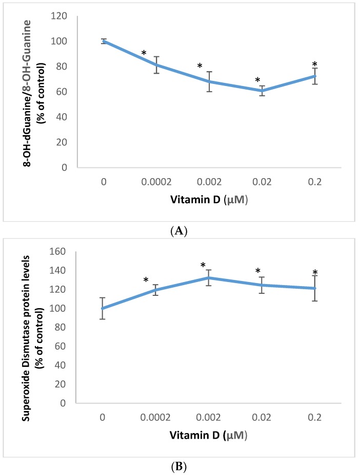
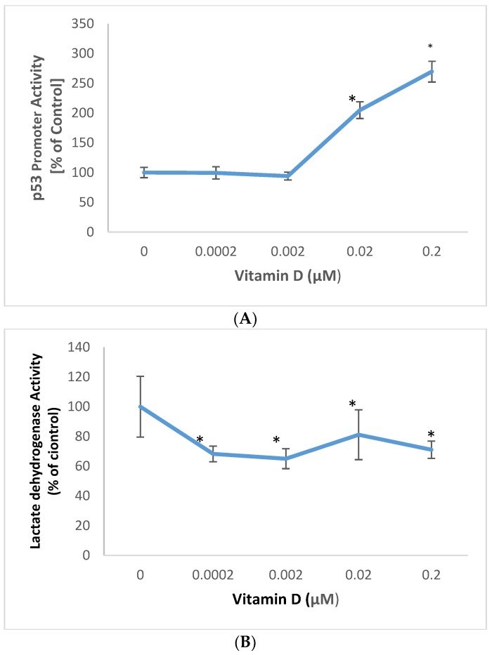
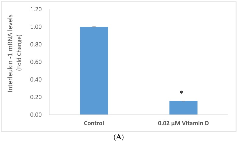
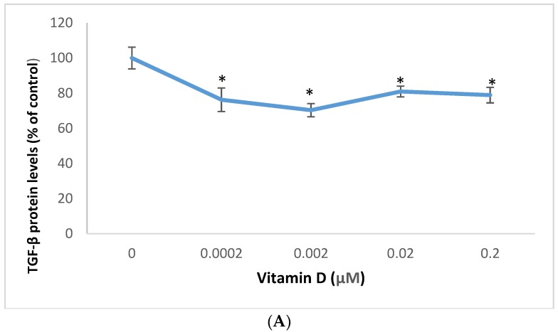
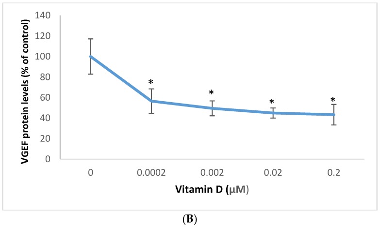
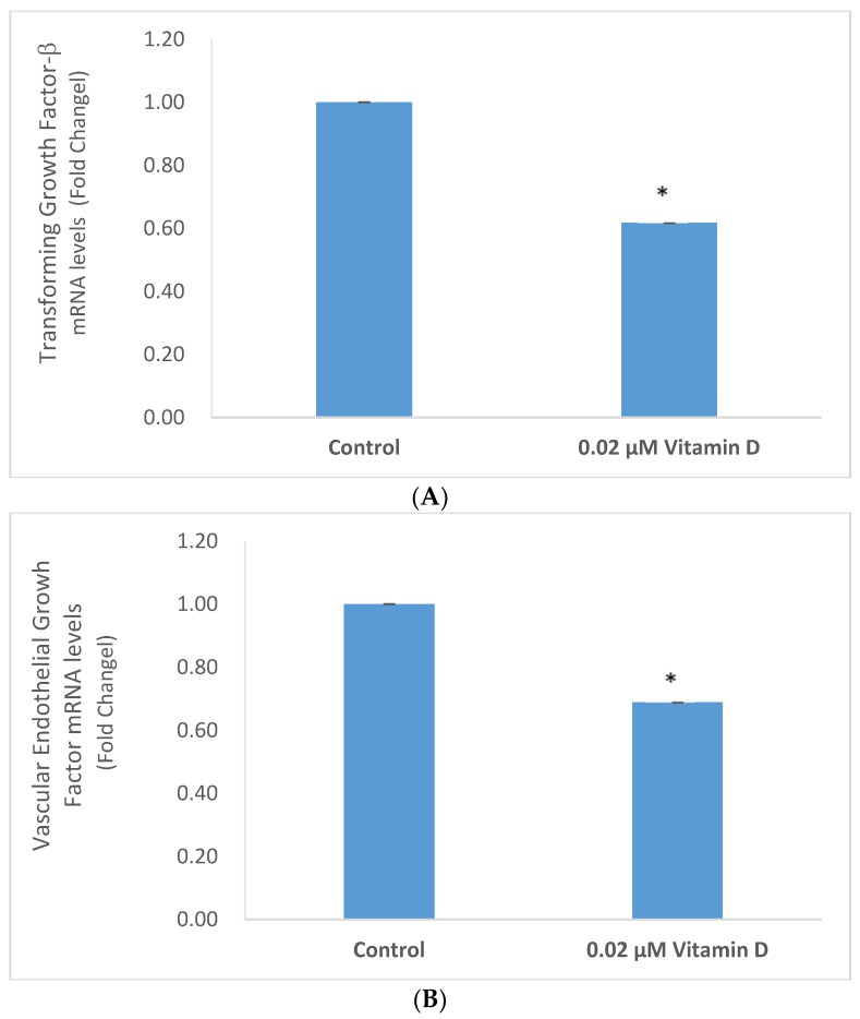
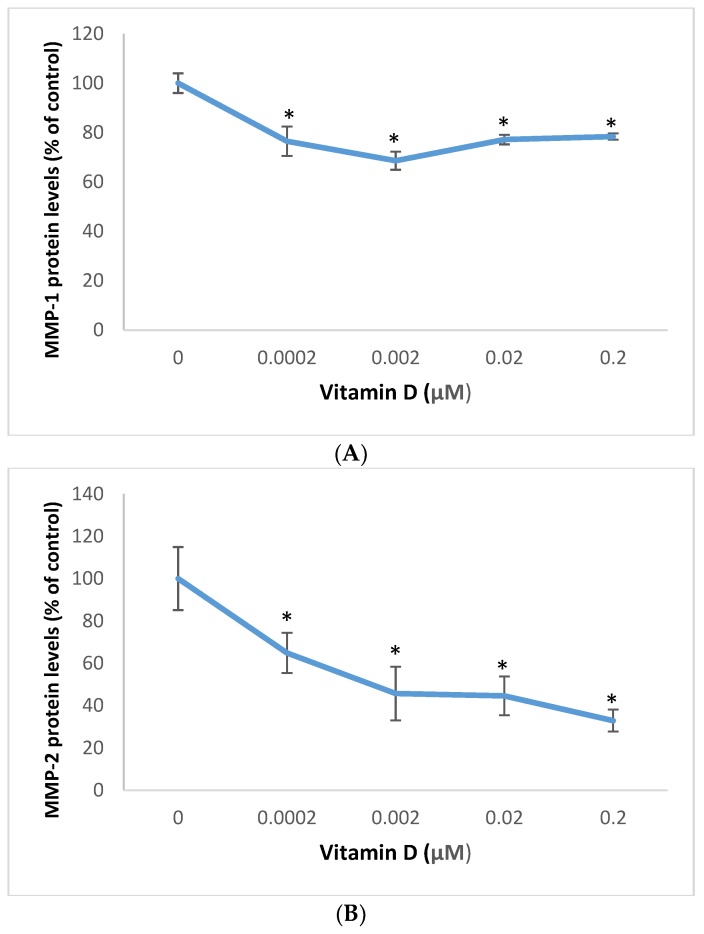
The causes of cancer include the cellular accumulation reactive oxygen species (ROS), which overrides the cellular antioxidants such as superoxide dismutase, from intrinsic aging, genetics, and exposure to environmental pollutants and ultraviolet (UV) radiation. The ROS damage biomolecules such as DNA (including p53 gene), RNA, and lipids, and activate inflammatory, angiogenic, and extracellular matrix (ECM) remodeling proteins; which collectively facilitate carcinogenesis. The 1α,25-dihydroxyvitamin D3 (Vitamin D) has anti-carcinogenic potential from its antioxidant, anti-inflammatory, and endocrine properties. We examined the anti-carcinogenic mechanism of vitamin D through the beneficial regulation of oxidative stress effects (oxidative DNA/RNA damage, superoxide dismutase expression, membrane damage, and p53 promoter activity), and expression (at the protein, mRNA and/or promoter levels) of inflammatory mediators (interleukin-1 (IL-1) and tumor necrosis factor-α (TNF-α)), angiogenic mediators (transforming growth factor-β (TGF-β), and vascular endothelial growth factor (VEGF)), and the ECM remodeling proteins (matrix metalloproteinases (MMP)-1 and MMP-2) by vitamin D in melanoma cells. Vitamin D inhibited oxidative DNA/RNA damage and membrane damage; and stimulated superoxide dismutase expression and p53 promoter activity in melanoma cells. It inhibited the expression of IL-1, TNF-α, TGF-β, VEGF, MMP-1 and MMP-2 by transcriptional or post-transcriptional mechanisms. We conclude that vitamin D is beneficial to melanoma cells through the inhibition of oxidative DNA/RNA damage, membrane damage, and the expression of inflammatory, angiogenic and ECM remodeling proteins; and the stimulation of superoxide dismutase expression and p53 promoter activity.
Keywords: interleukin-1; matrix metalloproteinases; oxidative DNA/RNA damage; p53; superoxide dismutase; transforming growth factor-β; tumor necrosis factor-α; vascular endothelial growth factor.
Conflict of interest statement
The authors declare no conflict of interest.
Figures

Similar articles
-
Beneficial regulation of matrixmetalloproteinases and their inhibitors, fibrillar collagens and transforming growth factor-beta by Polypodium leucotomos, directly or in dermal fibroblasts, ultraviolet radiated fibroblasts, and melanoma cells.Arch Dermatol Res. 2009 Aug;301(7):487-95. doi: 10.1007/s00403-009-0950-x. Epub 2009 Apr 17. Arch Dermatol Res. 2009. PMID: 19373483
-
Protective effects of 1,25 dihydroxyvitamin D3 and its analogs on ultraviolet radiation-induced oxidative stress: a review.Redox Rep. 2020 Dec;25(1):11-16. doi: 10.1080/13510002.2020.1731261. Redox Rep. 2020. PMID: 32093585 Free PMC article. Review.
-
Mesenchymal Stem Cells Modified with Heme Oxygenase-1 Have Enhanced Paracrine Function and Attenuate Lipopolysaccharide-Induced Inflammatory and Oxidative Damage in Pulmonary Microvascular Endothelial Cells.Cell Physiol Biochem. 2018;49(1):101-122. doi: 10.1159/000492847. Epub 2018 Aug 28. Cell Physiol Biochem. 2018. PMID: 30153667
-
Regulation of the extracellular matrix remodeling by lutein in dermal fibroblasts, melanoma cells, and ultraviolet radiation exposed fibroblasts.Arch Dermatol Res. 2007 Oct;299(8):373-9. doi: 10.1007/s00403-007-0779-0. Epub 2007 Aug 21. Arch Dermatol Res. 2007. PMID: 17710425
-
Cooperation of liver cells in health and disease.Adv Anat Embryol Cell Biol. 2001;161:III-XIII, 1-151. doi: 10.1007/978-3-642-56553-3. Adv Anat Embryol Cell Biol. 2001. PMID: 11729749 Review.
Cited by
-
Malignant Melanoma: An Overview, New Perspectives, and Vitamin D Signaling.Cancers (Basel). 2024 Jun 18;16(12):2262. doi: 10.3390/cancers16122262. Cancers (Basel). 2024. PMID: 38927967 Free PMC article. Review.
-
Vitamin D in Melanoma: Potential Role of Cytochrome P450 Enzymes.Life (Basel). 2024 Apr 15;14(4):510. doi: 10.3390/life14040510. Life (Basel). 2024. PMID: 38672780 Free PMC article. Review.
-
Genome-Protecting Compounds as Potential Geroprotectors.Int J Mol Sci. 2020 Jun 24;21(12):4484. doi: 10.3390/ijms21124484. Int J Mol Sci. 2020. PMID: 32599754 Free PMC article. Review.
References
MeSH terms
Substances
LinkOut - more resources
Full Text Sources
Medical
Research Materials
Miscellaneous
