Review
. 2020 Mar 5;21(5):1784.
doi: 10.3390/ijms21051784.

New Perspectives Related to the Bioluminescent System in Dinoflagellates: Pyrocystis lunula, a Case Study

Affiliations
Review

New Perspectives Related to the Bioluminescent System in Dinoflagellates: Pyrocystis lunula, a Case Study

Carlos Fajardo et al. Int J Mol Sci. .

Abstract

Pyrocystis lunula is considered a model organism due to its bioluminescence capacity linked to circadian rhythms. The mechanisms underlying the bioluminescent phenomenon have been well characterized in dinoflagellates; however, there are still some aspects that remain an enigma. Such is the case of the presence and diversity of the luciferin-binding protein (LBP), as well as the synthesis process of luciferin. Here we carry out a review of the literature in relation to the molecular players responsible for bioluminescence in dinoflagellates, with particular interest in P. lunula. We also carried out a phylogenetic analysis of the conservation of protein sequence, structure and evolutionary pattern of these key players. The basic structure of the luciferase (LCF) is quite conserved among the sequences reported to date for dinoflagellate species, but not in the case of the LBP, which has proven to be more variable in terms of sequence and structure. In the case of luciferin, its synthesis has been shown to be complex process with more than one metabolic pathway involved. The glutathione S-transferase (GST) and the P630 or blue compound, seem to be involved in this process. In the same way, various hypotheses regarding the role of bioluminescence in dinoflagellates are exposed.

Keywords: P630; blue compound; glutathione S-transferase; luciferase; luciferin; luciferin-binding protein.


Conflict of interest statement

The authors declare no conflict of interest.

Figures

Figure 1
Figure 1
Bioluminescence model in dinoflagellates, showing the effect of pH on both LBP and LCF. Modified from the proposed model by Rüdiger Hardeland (http://tolweb.org/notes/?note_id=5621) and work published by Morse et al. [30]. The structure of LBP is not known, the one shown here it was obtained by using the P. lunula sequence into Phyre2 and it is shown for illustration purposes only. The structure of LCF shown here was predicted in Phyre2 using the P. lunula sequence, as explain in the text and in Figure 2.
Figure 2
Figure 2
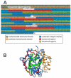
(A) Sequence of P. lunula LCF protein A (GenBank AAL40676) showing the Luciferase/LBP N-terminal, helical bundle and catalytic domains, as well as the two conserved regions within the catalytic domain. (B) Our prediction of the 2D/3D structure of the first helical bundle (yellow) and first catalytic domain of the LCF (blue) of P. lunula (GenBank AAL40676), showing the conserved regions 1 (red) and 2 (green), made with Phyre2 web server and visualized with EzMol, as described in the text.
Figure 3
Figure 3
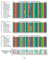
Sequence alignment of the conserved regions 1 (A) and 2 (B) found in the LCF comparing across the three catalytic domains and the available sequences from the species of the genera Alexandrium, Lingulodinium, Pyrocystis and Protoceratium. Numbers on top of each region correspond to the amino acid position in P. lunula protein (GenBank AAL40676). Asterisks are showing the conserved amino acids in each position. Coloring of the amino acids was made according to the same pattern displayed in MEGA7 software. Sequence logo was made using WebLogo, as described in the text.
Figure 3
Figure 3
Sequence alignment of the conserved regions 1 (A) and 2 (B) found in the LCF comparing across the three catalytic domains and the available sequences from the species of the genera Alexandrium, Lingulodinium, Pyrocystis and Protoceratium. Numbers on top of each region correspond to the amino acid position in P. lunula protein (GenBank AAL40676). Asterisks are showing the conserved amino acids in each position. Coloring of the amino acids was made according to the same pattern displayed in MEGA7 software. Sequence logo was made using WebLogo, as described in the text.
Figure 4
Figure 4
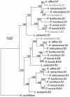
Molecular Phylogenetic analysis by a Bayesian method, showing the evolutionary pattern of the three catalytic domains in the species of dinoflagellates with protein sequences available, using the sole catalytic domain from N. scintillans as outgroup. The percentage of trees in which the associated taxa clustered together is shown next to the branches. There was a total of 297 positions in the final dataset and the tree is drawn to scale, with branch lengths measured in the number of substitutions per site.
Figure 5
Figure 5
Phylogenetic tree of Gonyaulacales among the dinoflagellates inferred from the three rDNA, two mitochondrial and four nuclear protein genes, modified from Orr et al. (2012). The tree is reconstructed with Bayesian inference (MrBayes) and included the different groups within dinoflagellates, but here only the Gonyaulacales are shown. Species in gray were not included in the original analysis by (Orr et al. 2012). The table is showing which of these species have reported LCF, LBP and GST protein sequences.
Figure 6
Figure 6
Sequence alignment of the three LCFs helical bundle domains contained in species of the genera Alexandrium, Lingulodinium, Ceratocorys, Pyrocystis and Protoceratium that have their protein published in GenBank. Coloring of the amino acids was made according to the same pattern displayed in MEGA7 software. Sequence logo was made using WebLogo, as described in the text.
Figure 7
Figure 7
Molecular Phylogenetic analysis by a Bayesian method, showing the evolutionary pattern of LBP from the species of the genera Alexandrium, Lingulodinium, Pyrocystis and Protoceratium that have their protein published in GenBank, using the sequence from N. scintillans as outgroup. The percentage of trees in which the associated taxa clustered together is shown next to the branches. There was a total of 435 positions in the final dataset and the tree is drawn to scale, with branch lengths measured in the number of substitutions per site.
Figure 8
Figure 8
Sequence alignment of the luciferase/LBP N terminal domain contained in the LCF, LBP and GST from the species of the genera Alexandrium, Lingulodinium, Pyrocystis and Protoceratium that have their protein published in GenBank. Coloring of the amino acids was made according to the same pattern displayed in MEGA7 software. Sequence logo was made using WebLogo, as described in the text.
Figure 9
Figure 9
Structure of the dinoflagellate luciferin, according to Wang and Liu, (2017).
Figure 10
Figure 10
Light emission process proposed by Fresneau et al. (1986) which is controlled by at least two successive reactions, where in the first the reduction of the luciferin precursor P630 is carried out by a NAD(P)H-dependent reductase, maybe GST, and in the second, the emission of light is carried out by LCF.

Similar articles

Cited by

References

    1. Widder E. Marine bioluminescence. Biosci. Explain. 2001;1:1–9.
    1. Haddock S., Moline M., Case J. Bioluminescence in the sea. Annu Rev. Mar. Sci. 2010;2:443–493. doi: 10.1146/annurev-marine-120308-081028. - DOI - PubMed
    1. Fuller C.W., Kreiss P., Seliger H.H. Particulate Bioluininescence in Dinoflagellates: Dissociation and Partial Reconstitution. Science. 1972;177:884–885. doi: 10.1126/science.177.4052.884. - DOI - PubMed
    1. Li L., Hastings J.W. The structure and organization of the luciferase gene in the photosynthetic dinoflagellate Gonyaulax polyedra. Plant. Mol. Biol. 1998;36:275–284. doi: 10.1023/A:1005941421474. - DOI - PubMed
    1. Shimomura O. Bioluminescence: Chemical Principles and Methods. World Scientific Publishing Co. Pte. Ltd.; Singapore: 2012. Revised Edition.