. 2020 Mar 5;12(3):602.
doi: 10.3390/cancers12030602.

Comprehensive Immunoprofiles of Renal Cell Carcinoma Subtypes

Affiliations

Comprehensive Immunoprofiles of Renal Cell Carcinoma Subtypes

Moonsik Kim et al. Cancers (Basel). .

Abstract

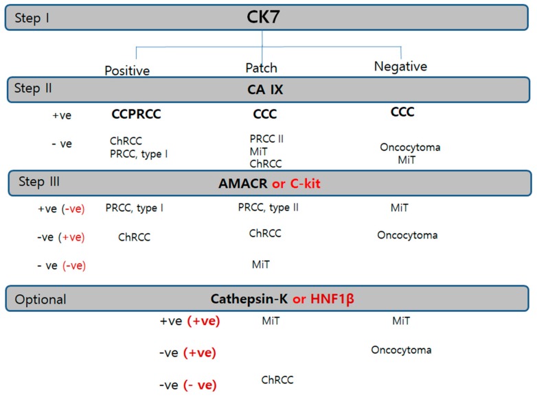
In recent years, renal epithelial tumors have been among the fastest reclassifying tumors, requiring updates to the tumor classification system. Nonetheless, immunohistochemistry (IHC) remains the most widely used tool for renal epithelial tumors. In this proposal, we aimed to create the most efficient IHC panel for categorizing the diverse subtypes of renal tumors, and to find out more specific immunohistochemical results in each subtype or each antibody. A total of 214 renal tumors were analyzed using 10 possible IHC markers to differentiate subtypes, including three major renal cell carcinoma (RCC) subtypes, clear-cell type (50 cases), papillary type (50 cases), and chromophobe type (20 cases), and minor subtypes (MiT RCC, 13 cases; collecting duct carcinoma, 5 cases; and oncocytoma, 10 cases). A triple immunomarker (cytokeratin 7 (CK7)-carbonic anhydrase IX (CAIX)- alpha-methylacyl-CoA racemase (AMACR)) panel is useful in particular high-grade clear-cell tumors. If IHC remains ambiguous, the use of an adjunctive panel can be suggested, including CD10, epithelial membrane antigen, cathepsin K, c-kit, hepatocyte nuclear factor 1-β, and E-cadherin. For an efficient immunohistochemical strategy for subtyping of RCC, we conclude that the CK7-CAIX-AMACR panel is the best primary choice for screening subtyping.

Keywords: Renal cell carcinoma; algorithm; diagnosis; immunohistochemistry; subtyping.


Conflict of interest statement

The authors declare that they have no conflict of interest.

Figures

Figure 1
Figure 1
Classic immunopanels for clear-cell renal cell carcinoma (CCRCC). (A1,A2) cytokeratin 7 labeling is mostly negative but exceptionally positive in macrocystic tubules (arrow). (B1,B2) Carbonic anhydrase IX (CAIX) is always positive as a box pattern, which is a pathognomonic sign. (C1,C2) CAIX labeling tends to be stronger and more diffuse in higher grades (grade 3–4) than in lower ones (grade 1–2). (D1,D2) Alpha-methylacyl-CoA racemase is variably positive but less intense than in papillary renal cell carcinoma (PRCC). Adjuvant immunopanels of CCRCC. (E1,E2) In CCRCC, immunoreactivities of epithelial membrane antigen and CD10 are closely correlated. EMA is more exaggerated (E1) and CD10 expression occurs in a saw-tooth luminal pattern (E2). (F1,F2) EMA (F1) labeling recapitulates exaggerated CD10 (F2) expression. (G1–G3) E-cadherin expression is frequently lost (G1,G2) or occasionally broken (arrows) (G3). Scale bars = 1 mm.
Figure 2
Figure 2
Papillary renal cell carcinoma (PRCC), type I. (A1,A2) Uniform structure predominantly composed of small slit-like tubules and luminal branched papillae is a characteristic of PRCC, type I. (BE) PRCC type I is characteristically cytokeratin 7 (CK7)++ (B)/Alpha-methylacyl-CoA racemase (AMACR)++ (C)/CD10− (D)/carbonic anhydrase IX (CAIX)− (E). PRCC, type II. (F1,F2) PRCC type II differs from type I in larger and more complicated papillae line by taller larger cells. Aggregates of foamy histiocytes in the stroma of papillae are frequently noted. (GK) Immunohistochemical (IHC) characteristic of PRCC, type II. PRCC, type II is usually CK7 negative (G), CAIX negative (H), epithelial membrane antigen+ (I) AMACR++ (J), and CD10++ (K1,K2). CD10 is basally distributed with a central sparing clubbing pattern (K2). Scale bars = 1 mm.
Figure 3
Figure 3
Clear-cell papillary renal cell carcinoma. (A1A4) In clear-cell papillary RCC (CCPRCC), histological findings include multilobulated small masses (A1) entirely composed of well-differentiated simple tubular structures mimicking distal tubules (A2) with intraluminal tufts (A3) and short papillary fronds in a glomeruloid pattern (A4). (BD) Immunohistochemical profiles. Cytokeratin 7 is diffusely and strongly positive (B), Carbonic anhydrase IX is positive with cup-shaped pattern, (C), Alpha-methylacyl-CoA racemase is generally negative except for macrocystic spaces (D), and CD10 as a saw-tooth luminal pattern (E). Scale bars = 1 mm.
Figure 4
Figure 4
Acquired cystic disease-associated renal cell carcinoma (ACD-RCC). (A1A3) In ACD-RCC, characteristic feature includes lace pattern (A1) with intratumoral oxalate crystals (A2) and coalescent vacuoles (A3). (B1B3) Immunoprofiles of ACD-RCC. Similar to papillary renal cell carcinoma type II, ACD-RCC demonstrates high alpha-methylacyl-CoA racemase (AMACR; B1) and CD10 (B2) expression with cytokeratin 7 negativity (B3). Immunohistochemistry is needed to differentiate between tubulocystic carcinoma (TCC) and multilocular cystic renal neoplasms. TCC is partly cystic (C1) and partly tubular (C2), which is characteristically AMACR+ (C3) and carbonic anhydrase IX (CAIX)-negative (C4) tumor, whereas multilocular cystic RCC shows hemorrhage-filled cyst (D1) and heaped-up profiles of clear cells (D2), and inverse immunoprofiles to TCC, AMACR-negative (D3), and CAIX-positive (D4) findings. Scale bars = 1 mm.
Figure 5
Figure 5
Microphthalmos translocation family RCC (MiT RCC). (A1A4) In MiT RCC, characteristic findings include intracystic papillary masses composed of clear cells (A2) with psammoma bodies (A1) and fibrins (A3) and sometimes marked reverse polarity (A4). However, these findings are not specific to diagnose MiT RCC by histology alone. (B,C) When MiT RCC is suspected, adequate immunohistochemical panel should be applied, beginning with cathepsin K staining (C1). Conventional markers such as CK7 (not shown), epithelial membrane antigen (B1), alpha-methylacyl-CoA racemase (B2), and carbonic anhydrase IX (not shown) are generally negative, while HMB45 (B3) and melan A (B4) are occasionally positive. A more definitive diagnosis can be made with transcription factor E3 (C2) or fluorescent in situ hybridization break-apart signals (C3). (D1D5) The characteristic findings transcription factor EB (TFEB)-related MiT RCC include nested fluoret sign (D1,D2), ballooning clear cells (D3), stromal hyalinization, fibrinous occlusive blood vessels with extensive peliosis-like changes (D4), and melanin pigment deposit in tumor cells (D5). (E1E4) Immunohistochemical profiles. TFEB (E1,E2), cathepsin K (E3), HMB45 (E4), paired box gene 8 (E5) are specifically positive. Scale bars = 1 mm.
Figure 6
Figure 6
Comparison of chromophobe renal cell carcinoma (ChRCC) with renal oncocytoma (RO). Differential diagnosis between these entities is difficult since they overlap in origin and morphology. (A1A4) cytokeratin 7 is highly intense and diffusely stained in ChRCC in contrast to RO showing perfect negativity. (B1B3) CD10 is generally negative, but focally patch positive, which is ignored. (C1C3) Alpha-methylacyl-CoA racemase is negative or patch positive. Epithelial membrane antigen (B4) and E-cadherin (C4) are diffusely positive both in ChRCC and RO. (D1D4) c-kit staining is basically positive in both entities. (E1E4) Hepatocyte nuclear factor-1β (HNF1β) staining is a highly useful immunomarker to rule out the possibility of ChRCC. Scale bars = 1 mm.
Figure 7
Figure 7
Alteration of immunoprofiles in aggressive chromophobe renal cell carcinoma (ChRCC). (A1–A3) Aggressive behavior is uncommon in ChRCC, but few cases show metastasis and even rupture intestine (A1). Tumor cells lost the nature of ChRCC, showing sarcomatoid differentiation (A2) and marked anaplasia (A3) corresponding to International Society of Urological Pathology grade 4. (B1–B4) Altered immunoprofiles. The most remarkable findings are aberrant expression of CD10 (B1) and alpha-methylacyl-CoA racemase (B2) with cytokeratin 7 (B3) and c-kit (B4) preservation. Scale bars = 1 mm.
Figure 8
Figure 8
Diagnostic immunohistochemistry algorithm based on the priority or sequence of trial in RCC. Cytokeratin 7 is the first trial universal marker in any kind of renal tumor. Carbonic anhydrase IX is the second trial cardinal marker. Alpha-methylacyl-CoA racemase or c-kit is used as the third panel to be applied in tubular/papillary or solid tumor, respectively. Cathepsin K or hepatocyte nuclear factor-1β is the subsequently used trial for confirmation of MiT family RCC or ChRCC. Abbreviations: CCC, clear-cell carcinoma; ChRCC, chromophobe renal cell carcinoma; CCPRCC, clear-cell papillary renal cell carcinoma; RO, renal oncocytoma; PRCC, papillary renal cell carcinoma; ACD-RCC, acquired cystic disease associated renal cell carcinoma; MiT RCC, microphthalmos gene translocation-associated renal cell carcinoma.

Similar articles

Cited by

References

    1. Inamura K. Renal Cell Tumors: Understanding Their Molecular Pathological Epidemiology and the 2016 WHO Classification. Int. J. Mol. Sci. 2017;18:2195. doi: 10.3390/ijms18102195. - DOI - PMC - PubMed
    1. Lopez-Beltran A., Cheng L., Vidal A., Scarpelli M., Kirkali Z., Blanca A., Montironi R. Pathology of renal cell carcinoma: An update. Anal. Quant. Cytopathol. Histpathol. 2013;35:61–76. - PubMed
    1. Skinnider B.F., Amin M.B. An immunohistochemical approach to the differential diagnosis of renal tumors. Semin. Diagn. Pathol. 2005;22:51–68. doi: 10.1053/j.semdp.2005.11.004. - DOI - PubMed
    1. Mikami S., Oya M., Mizuno R., Kosaka T., Ishida M., Kuroda N., Nagashima Y., Katsube K.I., Okada Y. Recent advances in renal cell carcinoma from a pathological point of view. Pathol. Int. 2016;66:481–490. doi: 10.1111/pin.12433. - DOI - PubMed
    1. Kuroda N., Tanaka A., Ohe C., Nagashima Y. Recent advances of immunohistochemistry for diagnosis of renal tumors. Pathol. Int. 2013;63:381–390. doi: 10.1111/pin.12080. - DOI - PubMed