Review
. 2020 Mar 5;12(3):233.
doi: 10.3390/pharmaceutics12030233.

Gene Therapy in Cancer Treatment: Why Go Nano?

Affiliations
Review

Gene Therapy in Cancer Treatment: Why Go Nano?

Catarina Roma-Rodrigues et al. Pharmaceutics. .

Abstract

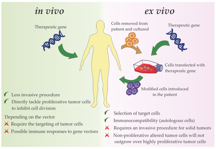
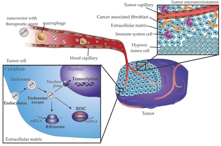
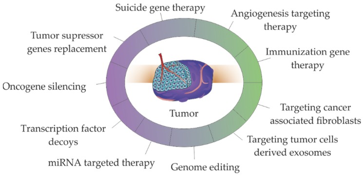
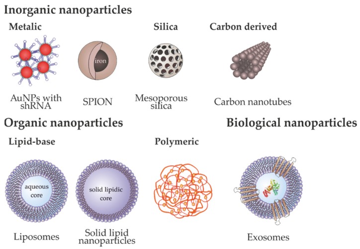
The proposal of gene therapy to tackle cancer development has been instrumental for the development of novel approaches and strategies to fight this disease, but the efficacy of the proposed strategies has still fallen short of delivering the full potential of gene therapy in the clinic. Despite the plethora of gene modulation approaches, e.g., gene silencing, antisense therapy, RNA interference, gene and genome editing, finding a way to efficiently deliver these effectors to the desired cell and tissue has been a challenge. Nanomedicine has put forward several innovative platforms to overcome this obstacle. Most of these platforms rely on the application of nanoscale structures, with particular focus on nanoparticles. Herein, we review the current trends on the use of nanoparticles designed for cancer gene therapy, including inorganic, organic, or biological (e.g., exosomes) variants, in clinical development and their progress towards clinical applications.

Keywords: gene delivery; gene therapy; nanomedicine; nanoparticles; tumor microenvironment.


Conflict of interest statement

vascular endothelial growth factor

Figures

Figure 1
Figure 1
Delivery strategies used for gene therapy directly targeting tumor cells or tumor microenvironment components, their major advantages (preceded by a green checkmark) and disadvantages (preceded by a red cross).
Figure 2
Figure 2
Barriers that nanoparticles must overcome for effective cancer gene delivery. In a systemic administration, nanoparticles should travel through the blood circulatory system, avoiding the immune system. The accumulation at the tumor occurs through passive targeting by the enhanced permeability and retention effect. Nanoparticles also have to penetrate into the most inaccessible areas of the tumor to reach the hypoxic tumor region with low oxygenation and dense extracellular matrix. After reaching tumor cells, nanoparticles should be internalized, which is mainly accomplished via endocytosis, and then escape from the endosome to efficiently deliver the cargo into the cytoplasm, when targeting RNA, or travel to the nucleus, when targeting DNA.
Figure 3
Figure 3
Major strategies used in non-viral gene therapies for cancer treatment. Therapies targeting the tumor microenvironment (in green), including angiogenesis targeting therapy, immunization gene therapy, targeting cancer associated fibroblasts and targeting tumor cells derived exosomes, also use the described molecular strategies (in purple), such as genes replacement, gene silencing, transcription factor decoys, miRNA targeted therapy and genome editing.
Figure 4
Figure 4
Nanoparticles used for gene delivery. Examples of metallic nanoparticles are gold nanoparticles (AuNPs) that can be functionalized with several molecules, e.g., short hairpin RNA (shRNA) for gene silencing. Other examples of inorganic nanoparticles are superparamagnetic iron oxide nanoparticles (SPION) containing an iron core coated with biocompatible polymers, mesoporous silica nanoparticles or carbon nanotubes. Examples of organic nanoparticles are polymeric nanoparticles, and liposomes and solid lipid nanoparticles (SLNs), which are lipid-base nanoparticles that differ mainly in the aqueous and lipidic core and the number of lipid layers. Exosomes are nanovesicles secreted by eukaryotic cells composed by a bi-lipidic membrane containing membrane proteins, that surround an aqueous lumen containing proteins and nucleic acids.

References

    1. World Health Organization. [(accessed on 28 November 2019)]; Available online: https://www.who.int/health-topics/cancer.
    1. Loponte S., Lovisa S., Deem A.K., Carugo A., Viale A. The many facets of tumor heterogeneity: Is metabolism lagging behind? Cancers. 2019;11:1574. doi: 10.3390/cancers11101574. - DOI - PMC - PubMed
    1. Hanahan D., Weinberg R.A. Hallmarks of cancer: The next generation. Cell. 2011;144:646–674. doi: 10.1016/j.cell.2011.02.013. - DOI - PubMed
    1. Wang M., Zhao J., Zhang L., Wei F., Lian Y., Wu Y., Gong Z., Zhang S., Zhou J., Cao K., et al. Role of tumor microenvironment in tumorigenesis. J. Cancer. 2017;8:761–773. doi: 10.7150/jca.17648. - DOI - PMC - PubMed
    1. Netea-Maier R.T., Smit J.W.A., Netea M.G. Metabolic changes in tumor cells and tumor-associated macrophages: A mutual relationship. Cancer Lett. 2018;413:102–109. doi: 10.1016/j.canlet.2017.10.037. - DOI - PubMed

LinkOut - more resources