. 2020 Mar 5;21(5):1795.
doi: 10.3390/ijms21051795.

Kynurenic Acid Protects Against Ischemia/Reperfusion-Induced Retinal Ganglion Cell Death in Mice

Affiliations

Kynurenic Acid Protects Against Ischemia/Reperfusion-Induced Retinal Ganglion Cell Death in Mice

Rooban B Nahomi et al. Int J Mol Sci. .

Abstract

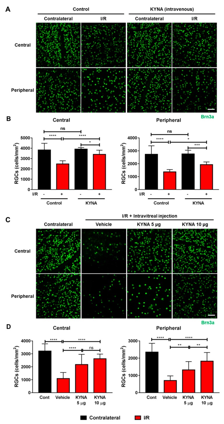
Background: Glaucoma is an optic neuropathy and involves the progressive degeneration of retinal ganglion cells (RGCs), which leads to blindness in patients. We investigated the role of the neuroprotective kynurenic acid (KYNA) in RGC death against retinal ischemia/reperfusion (I/R) injury.

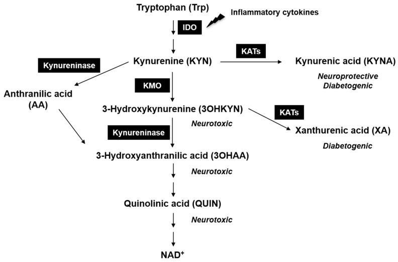
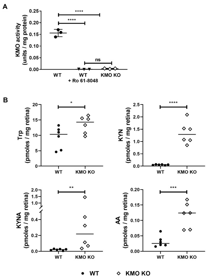
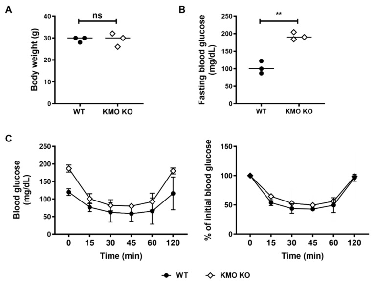
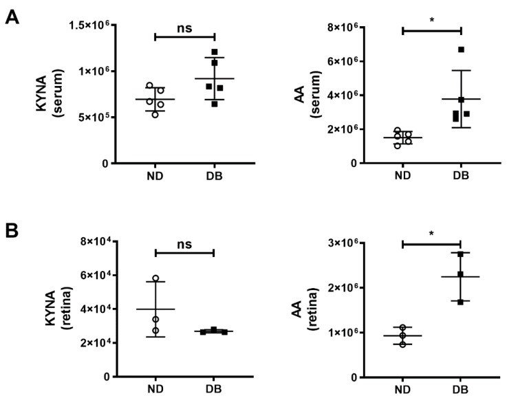
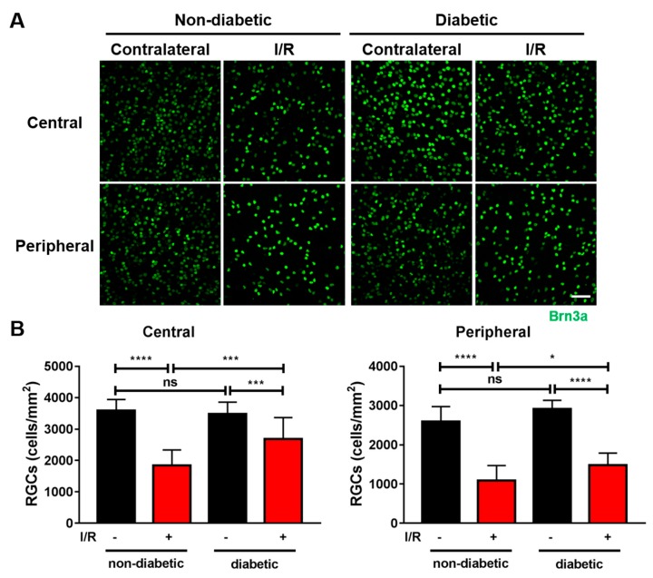
Methods: We injected KYNA intravenously or intravitreally to mice. We generated a knockout mouse strain of kynurenine 3-monooxygenase (KMO), an enzyme in the kynurenine pathway that produces neurotoxic 3-hydroxykynurenine. To test the effect of mild hyperglycemia on RGC protection, we used streptozotocin (STZ) induced diabetic mice. Retinal I/R injury was induced by increasing intraocular pressure for 60 min followed by reperfusion and RGC numbers were counted in the retinal flat mounts.

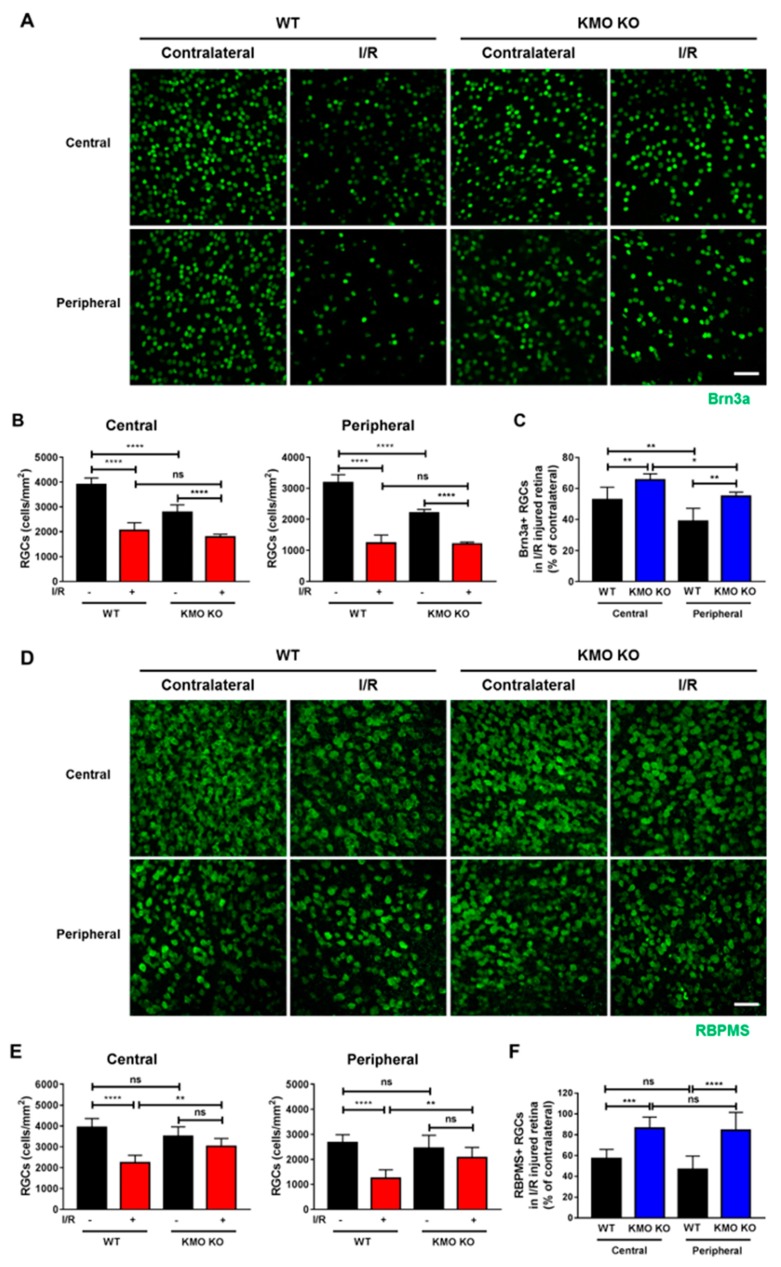
Results: Intravenous or intravitreal administration of KYNA protected RGCs against I/R injury. The I/R injury caused a greater loss of RGCs in wild type than in KMO knockout mice. KMO knockout mice had mildly higher levels of fasting blood glucose than wild type mice. Diabetic mice showed significantly lower loss of RGCs when compared with non-diabetic mice subjected to I/R injury.

Conclusion: Together, our study suggests that the absence of KMO protects RGCs against I/R injury, through mechanisms that likely involve higher levels of KYNA and glucose.

Keywords: diabetes; ischemia/reperfusion; kynurenic acid; kynurenine 3-monooxygenase; neuroprotection; retinal ganglion cells.


Conflict of interest statement

The authors declare no conflict of interest.

Figures

Figure 1
Figure 1
The kynurenine pathway (KP) and its metabolites. Indoleamine 2,3-dioxygenase (IDO) is the first enzyme in the KP. IDO synthesis is stimulated by inflammatory cytokines. Kynurenine 3-monooxygenase (KMO) converts kynurenine (KYN) to neurotoxic 3-hydroxykynurenine (3OHKYN). Quinolinic acid (QUIN), which is an N-methyl-D-aspartate (NMDA) receptor agonist, is also formed in this pathway. Kynurenine aminotransferases (KATs) convert KYN to neuroprotective kynurenic acid (KYNA) and kynureninase convert KYN to anthranilic acid (AA).
Figure 2
Figure 2
Retinal ganglion cell (RGC) loss in WT mice was blocked by intravenously and intravitreally injected KYNA. I/R injury was performed as described in the methods. Mice were intravenously injected with either vehicle alone or KYNA immediately after and 24 h after I/R injury (n = 5). Retinas were dissected out 14 days after I/R injury, flat mounted and immunostained for Brn3a (A,B) (n = 5). KYNA (5 μg (n = 8) or 10 μg (n = 7)) or vehicle alone was intravitreally injected immediately after I/R injury, and after 24 h. Retinas were dissected out after 14 days after I/R injury, flat mounted and immunostained for Brn3a (C,D) (n = 7–8), ns = not significant, * p < 0.05, ** p < 0.01, *** p < 0.001, and **** p < 0.0001. Scale bar = 50 μm.
Figure 3
Figure 3
KMO activity was present in wild type (WT) but not in KMO knockout (KO) mice, and serum KP metabolites were altered in KMO KO mice. KMO activity was measured in livers isolated from WT and KMO KO mice (A). The KMO activity in WT liver isolate was blocked by a KMO inhibitor Ro61-8048, confirming that the observed activity was due KMO (n = 3). Retinal KP metabolites were higher in the KMO KO than in WT mice (B). WT and KMO KO mice (n = 6) were euthanized and KP metabolites were determined by LC-MS/MS. ns = not significant, * p < 0.05, ** p < 0.01, *** p < 0.001, and **** p < 0.0001.
Figure 4
Figure 4
RGCs in KMO KO mice were protected from I/R injury. Retinas were dissected out 14 days after I/R injury, flat mounted and immunostained for Brn3a (AC) (n = 3) or RBPMS (DF) (WT; n = 4, KMO KO; n = 5). I/R injury reduced RGC numbers (central and peripheral retinas) in both WT and KMO KO mice. The percentage of Brn3a-positive and RBPMS-positive RGCs following I/R injury was higher in KMO KO mice compared to WT mice in both the central and peripheral retinas (C,F). The bar graphs represent the means ± SD of triplicate measurements. ns = not significant, * p < 0.05, ** p < 0.01, *** p < 0.001, and **** p < 0.0001. Scale bar = 50 μm. RBPMS = RNA-binding protein with multiple splicing.
Figure 5
Figure 5
Fasting blood glucose was higher in KMO KO mice than in WT mice, but body weights and insulin tolerance test results were similar. The body weights were similar in WT and KMO KO mice (A). Fasting blood glucose (FBG) was higher in KMO KO mice (B). The KMO KO mice responded to insulin like the WT mice (C). The line graphs represent the means ± SD of triplicate measurements. ns = not significant, ** p < 0.01.
Figure 6
Figure 6
The KYNA and AA were measured by LC-MS/MS (area under curve) in (A) serum (n = 5) and (B) retinas (n = 3) of non-diabetic (ND) and diabetic mice (DB). The KYNA levels in the serum and retina were not significantly different but the AA levels were higher in diabetic mice than in non-diabetic mice. The graphs represent the means ± SD of number of measurements mentioned above. ns = not significant and * p < 0.05.
Figure 7
Figure 7
Hyperglycemia inhibited RGC loss in mice subjected to I/R injury. I/R injury was performed in diabetic mice one-week after induction of diabetes. Retinas were dissected out 14 days after I/R injury, flat mounted and immunostained for Brn3a. (n = 4), ns = not significant, * p < 0.05, *** p < 0.001, and **** p < 0.0001. Scale bar = 50 μm.

References

    1. Quigley H.A., Broman A.T. The number of people with glaucoma worldwide in 2010 and 2020. Br. J. Ophthalmol. 2006;90:262–267. doi: 10.1136/bjo.2005.081224. - DOI - PMC - PubMed
    1. Gupta P., Zhao D., Guallar E., Ko F., Boland M.V., Friedman D.S. Prevalence of Glaucoma in the United States: The 2005–2008 National Health and Nutrition Examination Survey. Invest. Ophthalmol. Vis. Sci. 2016;57:2905–2913. doi: 10.1167/iovs.15-18469. - DOI - PMC - PubMed
    1. Almasieh M., Wilson A.M., Morquette B., Cueva Vargas J.L., Di Polo A. The molecular basis of retinal ganglion cell death in glaucoma. Prog. Retin. Eye Res. 2012;31:152–181. doi: 10.1016/j.preteyeres.2011.11.002. - DOI - PubMed
    1. Munemasa Y., Kitaoka Y. Molecular mechanisms of retinal ganglion cell degeneration in glaucoma and future prospects for cell body and axonal protection. Front. Cell Neurosci. 2012;6:60. doi: 10.3389/fncel.2012.00060. - DOI - PMC - PubMed
    1. Howell G.R., Soto I., Libby R.T., John S.W. Intrinsic axonal degeneration pathways are critical for glaucomatous damage. Exp. Neurol. 2013;246:54–61. doi: 10.1016/j.expneurol.2012.01.014. - DOI - PMC - PubMed

MeSH terms

Substances

LinkOut - more resources