. 2020 Mar 5;9(3):632.
doi: 10.3390/cells9030632.

MicroRNA-361-Mediated Inhibition of HSP90 Expression and EMT in Cervical Cancer Is Counteracted by Oncogenic lncRNA NEAT1

Affiliations

MicroRNA-361-Mediated Inhibition of HSP90 Expression and EMT in Cervical Cancer Is Counteracted by Oncogenic lncRNA NEAT1

Daozhi Xu et al. Cells. .

Abstract

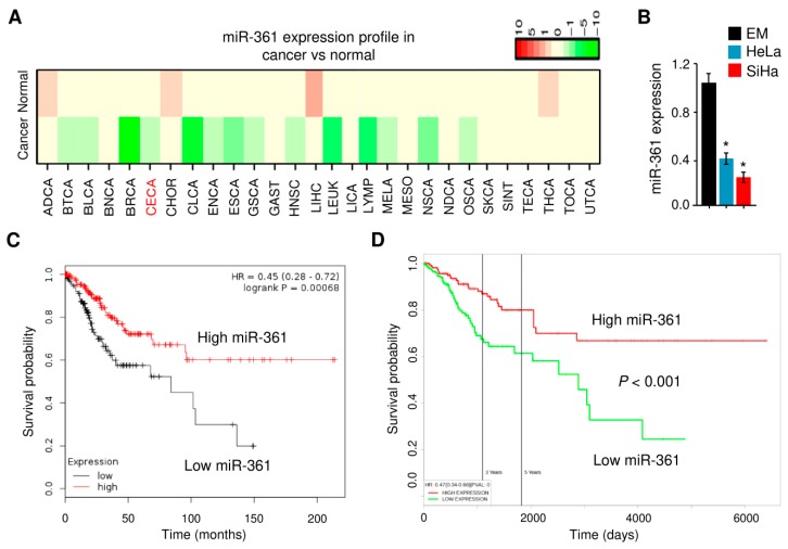
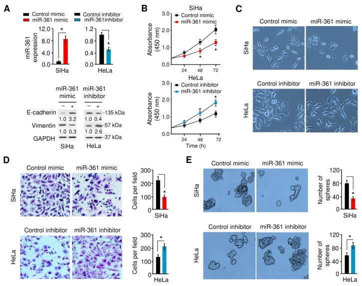
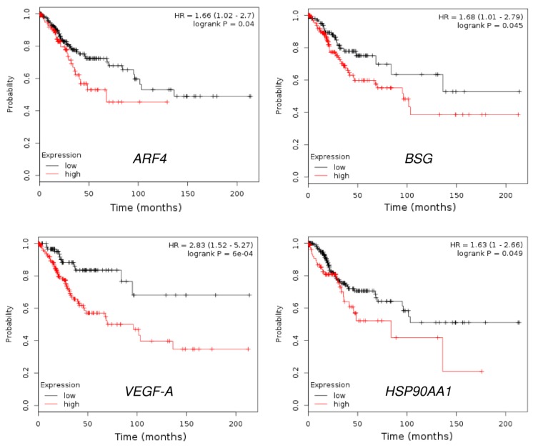
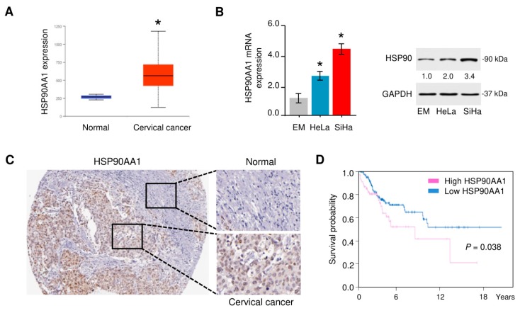
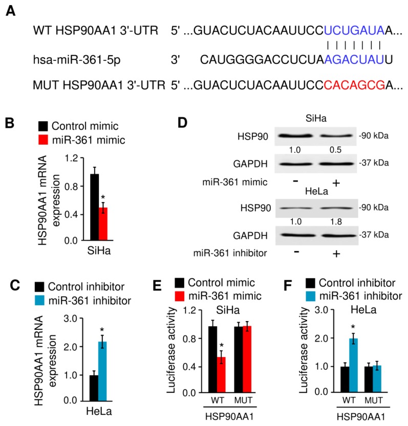
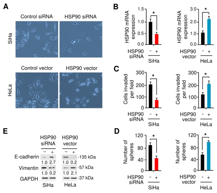
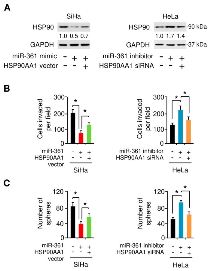
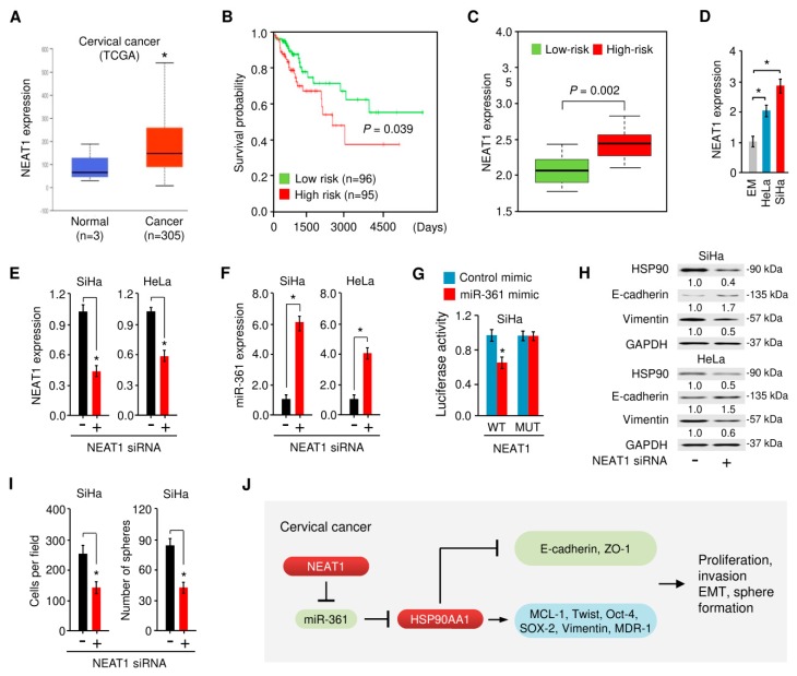
Epithelial-mesenchymal transition (EMT) is a key process contributing to cervical cancer (CC) metastasis, and microRNAs (miRNAs) modulate the expression of genes implicated in EMT. However, the accurate role of miR-361 in CC-associated EMT and the mechanisms underlying its function in CC remains largely unknown. The functional roles of miR-361 in CC cells were explored by a series of cell functional assays. Luciferase reporter assays were used to demonstrate the potential interaction between miR-361, HSP90, and long non-coding RNA (lncRNA) NEAT1. We detected a reduction of miR-361 expression in CC tissues compared with normal tissues, and miR-361 overexpression inhibited invasion and EMT phenotypes of CC cells by directly targeting a key EMT activator HSP90. Additionally, we detected significantly higher levels of HSP90 in CC tissues compared with normal tissues, and high expression of HSP90 predicted a poorer prognosis. We further identified NEAT1 as a significantly upregulated lncRNA in CC tissues and high expression of NEAT1 was associated with worse survival in CC patients. NEAT1 directly repressed miR-361 expression and played an oncogenic role in CC cell invasion and sphere formation. Conclusions: These results demonstrated that miR-361 directly targets HSP90 to inhibit the invasion and EMT features, and NEAT1 functions as an oncogenic lncRNA that suppresses miR-361 expression and induces EMT and sphere formation in CC cells, thus providing critical insights into the molecular pathways operating in this malignancy.

Keywords: EMT; HSP90AA1; NEAT1; cervical cancer; long non-coding RNA; microRNA-361.


Conflict of interest statement

The authors declare that there is no conflict of interest regarding the publication of this paper.

Figures

Figure 1
Figure 1
MiR-361 is aberrantly downregulated in cervical cancer tissues and cell lines. (A) Heatmap results showing differential expression patterns of miR-361 in different tumors and normal samples. MiR-361 expression in various types of cancer was searched in the dbDEMC v2.0 database. Green and red indicate downregulated and upregulated miR-361 expression in the corresponding cancer tissues, respectively. CECA: cervical cancer. (B) MiR-361 expression was measured in a human endometrial cell line (EM) and two human cervical cancer cell lines (HeLa and SiHa) using quantitative real-time PCR analysis. (C) The clinical significance of miR-361 expression in cervical cancer was evaluated by Kaplan–Meier survival analysis. The plots were generated using the KM-Plotter database. (D) The prognostic impact of miR-361 on cervical cancer was analyzed using the PROGmiR database. * p < 0.05.
Figure 2
Figure 2
MiR-361 inhibits epithelial-mesenchymal transition (EMT) and sphere formation in cervical cancer cells. (A) Upper panel: MiR-361 expression was examined in SiHa cells transfected with miR-361 mimic or control mimic, and in HeLa cells transfected with miR-361 inhibitor or control inhibitor using quantitative real-time PCR analysis. Lower panel: Representative Western blot images for E-cadherin, Vimentin, and GAPDH protein levels in cervical cancer cells after overexpression or knockdown of miR-361. Numbers denote fold change of the E-cadherin/GAPDH or Vimentin/GAPDH protein ratio in miR-361 mimic-transfected SiHa cells versus control mimic-transfected cells (set as default 1), or in miR-361 inhibitor-transfected HeLa cells versus control inhibitor-transfected cells (set as default 1). (B) Growth curves of cervical cancer cells transfected as indicated were determined using CCK-8 assays. (C) MiR-361 affects cervical cancer cell morphology. (D, E) The effects of miR-361 expression on cell invasion (D) and sphere formation (E) were assessed using the Matrigel invasion and sphere formation assays. * p < 0.05.
Figure 3
Figure 3
KEGG pathway and GO enrichment analysis for predicted miR-361 target genes using the DAVID database. (A) Enrichment analysis for KEGG pathways. (B) GO analysis according to the biological process.
Figure 4
Figure 4
Box plots representing the expression level of the predicted miR-361 target genes in cervical.cancer tissues and normal cervical tissues (Oncomine database). (A) ARF4 expression from Biewenga dataset, (B) DEPDC1B expression form Biewenga dataset, (C) EPHA4 expression from Scotto dataset, (D) ABI2 expression from Pyeon dataset, (E) BSG expression from Scotto dataset, (F) HSP90AA1 expression from Pyeon dataset, (G) OGDH expression from Biewenga dataset, (H) PHACTR4 expression from the TCGA dataset, (I) RAC1 expression from Scotto dataset, (J) TMEM18 expression from Biewenga dataset, (K) Twist1 expression from Scotto dataset, and (L) VEGF-A expression from Biewenga dataset.
Figure 5
Figure 5
The prognostic value of ARF4, BSG, VEGF-A, and HSP90 expression in cervical cancer (KM-Plotter database).
Figure 6
Figure 6
HSP90 is highly expressed in cervical cancer tissues. (A) The expression of HSP90 in cervical cancer tissues and normal tissues (UALCAN database). (B) Left panel: HSP90 expression was measured in a human endometrial cell line (EM) and two human cervical cancer cell lines (HeLa and SiHa) using quantitative real-time PCR analysis. Right panel: representative Western blot images for HSP90 and GAPDH protein levels in cervical cancer cell lines and normal EM cells. Numbers denote fold change of the HSP90/GAPDH protein ratio in cervical cancer cell lines versus normal EM cells (set as default 1). (C) HSP90 protein expression was upregulated in cervical cancer tissues compared with adjacent normal tissues (Human Protein Atlas database). (D) The prognostic value of HSP90 expression in cervical cancer (Human Protein Atlas database). * p < 0.05.
Figure 7
Figure 7
MiR-361 binds to HSP90 mRNA and suppresses its expression. (A) Illustration of the predicted binding site between miR-361 and HSP90 mRNA, with mutant sites shown in red. (B) The mRNA expression of HSP90 in SiHa cells transfected with miR-361 mimic or control mimic. (C) The mRNA level of HSP90 in HeLa cells transfected with miR-361 inhibitor and control inhibitor. (D) Western blot analysis of HSP90 and GAPDH expression in cervical cancer cells transfected as indicated. Numbers denote fold change of the HSP90/GAPDH protein ratio in miR-361 mimic-transfected SiHa cells versus control mimic-transfected cells (set as default 1), or in miR-361 inhibitor-transfected HeLa cells versus control inhibitor-transfected cells (set as default 1). (E) Luciferase assays in SiHa cells transfected with wild-type (WT) or mutant (MUT) HSP90 3′-UTR, together with miR-361 mimic or control mimic. (F) Luciferase assays in HeLa cells transfected with WT or MUT HSP90 3′-UTR, together with miR-361 inhibitor or control inhibitor. * p < 0.05.
Figure 8
Figure 8
HSP90 enhances EMT and sphere formation in cervical cancer cells. (A) The morphology of.SiHa and HeLa cells transfected as indicated are shown. (B) Quantitative real-time PCR analysis of HSP90 mRNA expression was examined in SiHa and HeLa cells transfected as indicted. (C,D) The impact of HSP90 expression on cell invasion (C) and sphere formation (D) were assessed using the Matrigel invasion assay and sphere formation assays. (E) Representative Western blot images for E-cadherin, Vimentin, and GAPDH protein levels in cervical cancer cells after overexpression or knockdown of HSP90. Numbers denote fold change of the E-cadherin/GAPDH or Vimentin/GAPDH protein ratio in HSP90 siRNA-transfected SiHa cells versus control siRNA-transfected cells (set as default 1), or in HSP90 vector-transfected HeLa cells versus control vector-transfected cells (set as default 1). * p < 0.05.
Figure 9
Figure 9
MiR-361 suppresses EMT and sphere formation in cervical cancer cells by inhibiting HSP90 expression. (A) HSP90 and GAPDH protein level in SiHa cells transfected with miR-361 mimic (or control mimic), together with (or without) HSP90 vector, and in HeLa cells transfected with miR-361 inhibitor (or control inhibitor), together with (or without) HSP90 siRNA. Numbers denote fold change of the HSP90/GAPDH protein ratio in cervical cancer cells transfected as indicated. (B,C) Cell invasion (B) and sphere formation (C) capacity was detected in cervical cancer cells transfected as indicated. * p < 0.05.
Figure 10
Figure 10
NEAT1-mediated miR-361 downregulation contributes to EMT and sphere formation of cervical cancer cells via increasing the HSP90 expression. (A) The expression of NEAT1 in cervical cancer tissues and normal tissues (UALCAN database). (B) The patients from the TCGA cervical cancer dataset in the SurvExpress database were divided into low- and high-risk groups and survival differences between the two groups were demonstrated with Kaplan-Meier survival curves. (C) The expression level of NEAT1 in low- and high-risk groups. (D) NEAT1 expression was examined in a normal endometrial cell line (EM) and in cervical cancer cell lines (HeLa and SiHa) using quantitative real-time PCR analysis. (E, F) NEAT1 (E) and miR-361 (F) expression in SiHa and HeLa cells transfected with NEAT1 siRNA or control siRNA. (G) Luciferase activities were measured in SiHa cells co-transfected with the reporter vectors (wild-type or mutant NEAT1), together with (or without) miR-361 mimic. (H) Western blot analysis of HSP90, E-cadherin, Vimentin, and GAPDH expression in SiHa and HeLa cells transfected with NEAT1 siRNA or control siRNA. Numbers denote fold change of the HSP90/GAPDH, E-cadherin/GAPDH or Vimentin/GAPDH protein ratio in NEAT1 siRNA-transfected cervical cancer cells versus control siRNA-transfected cells (set as default 1). (I) Cell invasion and sphere formation capacity were detected in SiHa cells transfected with NEAT1 siRNA or control siRNA. (J) A scheme illustrating the mechanism by which lncRNA NEAT1 sponges miR-361 to upregulate the expression of HSP90, thereby promoting EMT and cancer stem cell properties in cervical cancer cells. * p < 0.05.

References

    1. Li H., Wu X., Cheng X. Advances in diagnosis and treatment of metastatic cervical cancer. J. Gynecol. Oncol. 2016;27:e43. doi: 10.3802/jgo.2016.27.e43. - DOI - PMC - PubMed
    1. Valastyan S., Weinberg R.A. Tumor metastasis: Molecular insights and evolving paradigms. Cell. 2011;147:275–292. doi: 10.1016/j.cell.2011.09.024. - DOI - PMC - PubMed
    1. Konno Y., Dong P., Xiong Y., Suzuki F., Lu J., Cai M., Watari H., Mitamura T., Hosaka M., Hanley S.J., et al. MicroRNA-101 targets EZH2, MCL-1 and FOS to suppress proliferation, invasion and stem cell-like phenotype of aggressive endometrial cancer cells. Oncotarget. 2014;5:6049–6062. doi: 10.18632/oncotarget.2157. - DOI - PMC - PubMed
    1. Lee W.S., Kim N., Park Y.R., Oh H.H., Myung E., Kim S.H., Yu H.M., Kim M.Y., Oak C.Y., Chung C.Y., et al. Myeloid cell leukemia-1 promotes epithelial-mesenchymal transition of human gastric cancer cells. Oncol. Rep. 2015;34:1011–1016. doi: 10.3892/or.2015.4040. - DOI - PubMed
    1. Dai X., Ge J., Wang X., Qian X., Zhang C., Li X. OCT4 regulates epithelial-mesenchymal transition and its knockdown inhibits colorectal cancer cell migration and invasion. Oncol. Rep. 2013;29:155–160. doi: 10.3892/or.2012.2086. - DOI - PubMed

Publication types

MeSH terms