Review
. 2020 Apr;29(2):185-208.
doi: 10.1016/j.soc.2019.11.003.

Pathologic Considerations in Gastroenteropancreatic Neuroendocrine Tumors

Affiliations
Review

Pathologic Considerations in Gastroenteropancreatic Neuroendocrine Tumors

Andrew M Bellizzi. Surg Oncol Clin N Am. 2020 Apr.

Abstract

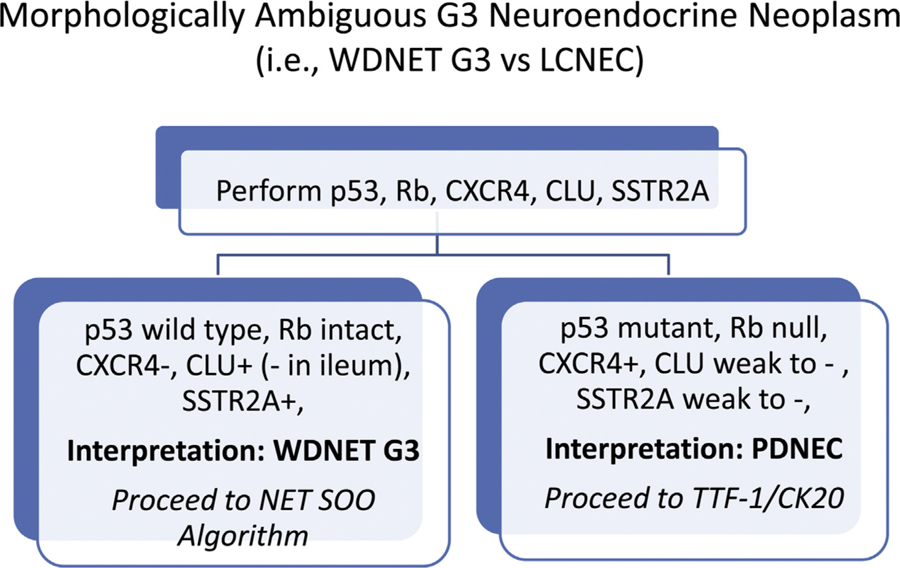
This review serves as a primer on contemporary neuroendocrine neoplasm classification, with an emphasis on gastroenteropancreatic well-differentiated neuroendocrine tumors. Topics discussed include general features of neuroendocrine neoplasms, general neuroendocrine marker immunohistochemistry, the distinction of well-differentiated neuroendocrine tumor from pheochromocytoma/paraganglioma and other diagnostic mimics and poorly differentiated neuroendocrine carcinoma from diagnostic mimics, the concepts of differentiation and grade and the application of Ki-67 immunohistochemistry to determine the latter, the various WHO classifications of neuroendocrine neoplasms including the 2019 WHO classification of gastroenteropancreatic tumors, organ-specific considerations for gastroenteropancreatic well-differentiated neuroendocrine tumors, immunohistochemistry to determine site of origin in metastatic well-differentiated neuroendocrine tumor of occult origin, immunohistochemistry in the distinction of well-differentiated neuroendocrine tumor G3 from large cell neuroendocrine carcinoma, and, finally, required and recommended reporting elements for biopsies and resections of gastroenteropancreatic neuroendocrine epithelial neoplasms.

Keywords: Differentiation; Grade; Immunohistochemistry; Ki-67; Neuroendocrine; Site of origin; WHO classification.


Conflict of interest statement

Disclosure This work was supported by NIH grant P50 CA174521-01A1 (AMB).

Figures

Fig. 1.
Fig. 1.
Neuroendocrine neoplasms. (A) Well-differentiated neuroendocrine tumor. (B) Paraganglioma. (C) Large cell neuroendocrine carcinoma. (D) Small cell neuroendocrine carcinoma.
Fig. 2.
Fig. 2.
Poorly differentiated neuroendocrine carcinoma versus nonneuroendocrine carcinoma with “occult” neuroendocrine differentiation. (A) This small cell lung cancer demonstrates (B) diffuse, strong synaptophysin expression. Poorly differentiated neuroendocrine carcinomas are often general neuroendocrine marker weak-to-negative, in which case dot-like keratin positivity, TTF-1 expression, and Rb loss may serve as surrogate markers for the presence of neuroendocrine differentiation. (C) This poorly differentiated lung adenocarcinoma was initially diagnosed as “poorly differentiated carcinoma with neuroendocrine features” based on (D) this synaptophysin immunostain. Ten to twenty percent of non-neuroendocrine carcinomas express general neuroendocrine markers, usually in weak, patchy fashion (as in this case), but occasionally quite strongly. This finding has no clear bearing on prognosis or therapy.
Fig. 3.
Fig. 3.
Paraganglioma/pheochromocytoma versus well-differentiated neuroendocrine tumor. While well-differentiated neuroendocrine tumors should be broad-spectrum keratinpositive, this (A) paraganglioma expressed (B) GATA-3, supporting the diagnosis. (C) Loss of SDHB expression (with intact signal in endothelium and sustentacular cells) raises the possibility of a hereditary tumor and is prognostically adverse.
Fig. 4.
Fig. 4.
Ki-67 proliferation index heterogeneity. (A) Low-power photomicrograph of Ki-67-immunostained well-differentiated neuroendocrine neoplasm highlights areas of (B) frequent (14% in this image) and (C) absent (0%) tumor cell staining. The Ki-67 proliferation index should be based on a count of at least 500 tumor nuclei in a “hotspot.”
Fig. 5.
Fig. 5.
Ki-67 proliferation index “Eyeball Estimate” versus manual count. This Ki-67 immunostain was originally reported as “approximately 30%” by the referring pathologist. A manual count of a camera-captured image found a proliferation index of 19.9% (175 Ki-67-immunostained tumor cells/878 tumor cells counted).
Fig. 6.
Fig. 6.
Well-differentiated neuroendocrine tumor G3. (A) Pancreatic well-differentiated neuroendocrine tumor with (B) Ki-67 proliferation index heterogeneity readily noted at low power. (C, D) Whereas most of the tumor had a low-to-intermediate proliferation index (3.5% in D), there was a distinct clone with a high proliferation index (44%) (E, F). This tumor pursued an aggressive clinical course with the patient dying 9 months after distal pancreatectomy.
Fig. 7.
Fig. 7.
University of Iowa immunohistochemical algorithm for well-differentiated neuroendocrine tumor site of origin.
Fig. 8.
Fig. 8.
Metastatic well-differentiated neuroendocrine tumor of unknown primary—site of origin immunohistochemistry. (A) This well-differentiated neuroendocrine tumor metastatic to liver expresses (B) CDX2 and not (C) islet 1 (depicted) or PAX6, supporting a midgut (ie, jejunoileal) origin. Subsequent Ga 68-DOTATATE scan demonstrated focal uptake in the distal ileum.
Fig. 9.
Fig. 9.
Simplified immunohisochemical algorithm for well-differentiated neuroendocrine tumor site of origin.
Fig. 10.
Fig. 10.
Immunohistochemical algorithm for morphologically ambiguous G3 neuroendocrine epithelial neoplasms.
Fig. 11.
Fig. 11.
p53 and Rb immunohistochemistry to distinguish well-differentiated neuroendocrine tumor G3 versus large cell neuroendocrine carcinoma. (A) This intermediate- to high-grade neuroendocrine epithelial neoplasm demonstrates (B) wild-type pattern p53 staining and (C) intact Rb expression, supporting the diagnosis of well-differentiated neuroendocrine tumor. (D) This high-grade neuroendocrine epithelial neoplasm demonstrates (E) missensemutation pattern p53 staining (ie, diffuse, strong staining obscuring nuclear detail) and (F) loss of Rb expression, supporting the diagnosis of large cell neuroendocrine carcinoma.

References

    1. Ionescu DN, Treaba D, Gilks CB, et al. Nonsmall cell lung carcinoma with neuroendocrine differentiation–an entity of no clinical or prognostic significance. Am J Surg Pathol 2007;31(1):26–32. - PubMed
    1. Howe MC, Chapman A, Kerr K, et al. Neuroendocrine differentiation in non-small cell lung cancer and its relation to prognosis and therapy. Histopathology 2005; 46(2):195–201. - PubMed
    1. Graziano SL, Tatum AH, Newman NB, et al. The prognostic significance of neuroendocrine markers and carcinoembryonic antigen in patients with resected stage I and II non-small cell lung cancer. Cancer Res 1994;54(11):2908–13. - PubMed
    1. Bellizzi AM. Assigning site of origin in metastatic neuroendocrine neoplasms: a clinically significant application of diagnostic immunohistochemistry. Adv Anat Pathol 2013;20(5):285–314. - PubMed
    1. Bellizzi A p53 and Rb immunohistochemistry as molecular surrogates show distinctive patterns in visceral and cutaneous poorly differentiated neuroendocrine carcinomas. Mod Pathol 2019;32(Supplement 2):540A.

Publication types

Substances

Supplementary concepts

LinkOut - more resources