Review
. 2020 May:121:38-46.
doi: 10.1016/j.molimm.2020.02.021. Epub 2020 Mar 6.

Transcriptional regulation of DC fate specification

Affiliations
Review

Transcriptional regulation of DC fate specification

Cédric Bosteels et al. Mol Immunol. 2020 May.

Abstract

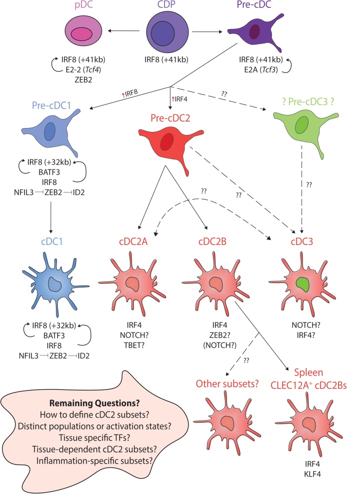
Dendritic cells function in the immune system to instruct adaptive immune cells to respond accordingly to different threats. While conventional dendritic cells can be subdivided into two main subtypes, termed cDC1s and cDC2s, it is clear that further heterogeneity exists within these subtypes, particularly for cDC2s. Understanding the signals involved in specifying each of these lineages and subtypes thereof is crucial to (i) enable us to determine their specific functions and (ii) put us in a position to be able to target these cells to promote or prevent a specific function in any given disease setting. Although we still have much to learn regarding the specification of these cells, here we review the most recent advances in our understanding of this and highlight some of the next questions for the future.

Keywords: DC fate; Dendritic cells; Irf4; Irf8; Klf4; Notch; Tbet; Transcription Factors; Zeb1; Zeb2.


Figures

Fig. 1
Fig. 1
Recent advances in cDC fate specification by Transcription Factors. Summary figure detailing recent advances in our understanding of transcriptional control of DC fate, highlighting remaining unknowns and main questions for the future. cDC1s develop from CDPs through pre-cDC1 intermediates. In mice, progression to cDC1 is achieved through increasing expression of IRF8 and switching from the +41 kb enhancer to the +32 kb enhancer at the pre-cDC stage. cDC1s express IRF8, BATF3, ID2, NFIL3 (red arrows) and absolutely require IRF8 (maintained through BATF3 and IRF8 autoactivation) for their development and maintenance. Expression of NFIL3, inhibits ZEB2 expression which in turn enables ID2 expression, normally repressed by ZEB2. Like cDC1s, cDC2s develop from CDPs but via a pre-cDC2 intermediate. There is still much to learn regarding cDC2s. Typically IRF4 is thought of as the main cDC2 TF in mice, however, it is not required for the development of all subsets, but may instead be important for survival and/or migration of specific subsets. In recent years, considerable heterogeneity has been demonstrated within the cDC2 population including the recent description of Tbet+ and Tbet cDC2s termed cDC2As and cDC2Bs respectively identified in mouse and human spleen. To date no distinct precursors for these subsets have been identified. In mice, cDC2As appear to also express and require NOTCH signaling being enriched for Rbpj and Runx binding sites. Whether Tbet is absolutely required for cDC2As remains to be studied. Some evidence suggests murine cDC2Bs (or a subset thereof), may require ZEB2 for their development but this remains to be directly investigated. Clec12A+ cDC2Bs, one subset of cDC2Bs, have been shown to require KLF4 in mice. Requirements for the other subsets remain to be studied. In human PBMCs, a pro-inflammatory cDC3 population has also recently been described, the TFs mediating this population and the precise progenitor remain to be identified however, they were shown to express higher NOTCH2 than KLF4. Thus the relationship of these cDC3s to cDC2As requires further investigation. Key questions for the future, especially related to heterogeneity within the cDC2 lineage, including understanding the role of the tissue microenvironment in cDC2 biology. How many distinct populations of cDC2s are there? Are there different subsets based on tissue? Do distinct TFs play a role in each tissue to generate the final cDC2 phenotype? Are cDC2 subsets arising from distinct developmental pathways or do they represent alternative activation states? Understanding this, will be a main goal in the coming years.

Similar articles

Cited by

References

    1. Ainsua-Enrich E., Hatipoglu I., Kadel S., Turner S., Paul J., Singh S., Bagavant H., Kovats S. IRF4-dependent dendritic cells regulate CD8+ T-cell differentiation and memory responses in influenza infection. Mucosal Immunol. 2019;12:1025–1037. doi: 10.1038/s41385-019-0173-1. - DOI - PMC - PubMed
    1. Akbari M., Honma K., Kimura D., Miyakoda M., Kimura K., Matsuyama T., Yui K. IRF4 in dendritic cells inhibits IL-12 production and controls Th1 immune responses against leishmania major. J. Immunol. 2014;192:2271–2279. doi: 10.4049/jimmunol.1301914. - DOI - PubMed
    1. Aliberti J., Schulz O., Pennington D.J., Tsujimura H., Reis e Sousa C., Ozato K., Sher A. Essential role for ICSBP in the in vivo development of murine CD8α+ dendritic cells. Blood. 2003;101:305–310. doi: 10.1182/blood-2002-04-1088. - DOI - PubMed
    1. Askenase M.H., Han S.-J., Byrd A.L., Morais da Fonseca D., Bouladoux N., Wilhelm C., Konkel J.E., Hand T.W., Lacerda-Queiroz N., Su X., Trinchieri G., Grainger J.R., Belkaid Y. Bone-Marrow-Resident nk cells prime monocytes for regulatory function during infection. Immunity. 2015;42:1130–1142. doi: 10.1016/j.immuni.2015.05.011. - DOI - PMC - PubMed
    1. Auderset F., Schuster S., Fasnacht N., Coutaz M., Charmoy M., Koch U., Favre S., Wilson A., Trottein F., Alexander J., Luther S.A., MacDonald H.R., Radtke F., Tacchini-Cottier F. Notch signaling regulates follicular helper t cell differentiation. J. Immunol. 2013;191:2344–2350. doi: 10.4049/jimmunol.1300643. - DOI - PubMed

Publication types