. 2020 Apr 15;204(8):2192-2202.
doi: 10.4049/jimmunol.1900912. Epub 2020 Mar 9.

Age-Dependent Effects of Type I and Type III IFNs in the Pathogenesis of Bordetella pertussis Infection and Disease

Affiliations

Age-Dependent Effects of Type I and Type III IFNs in the Pathogenesis of Bordetella pertussis Infection and Disease

Jeremy Ardanuy et al. J Immunol. .

Abstract

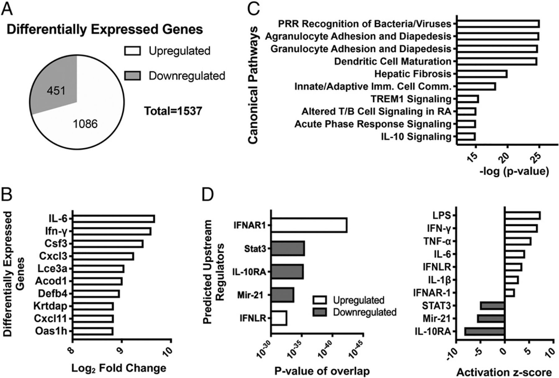
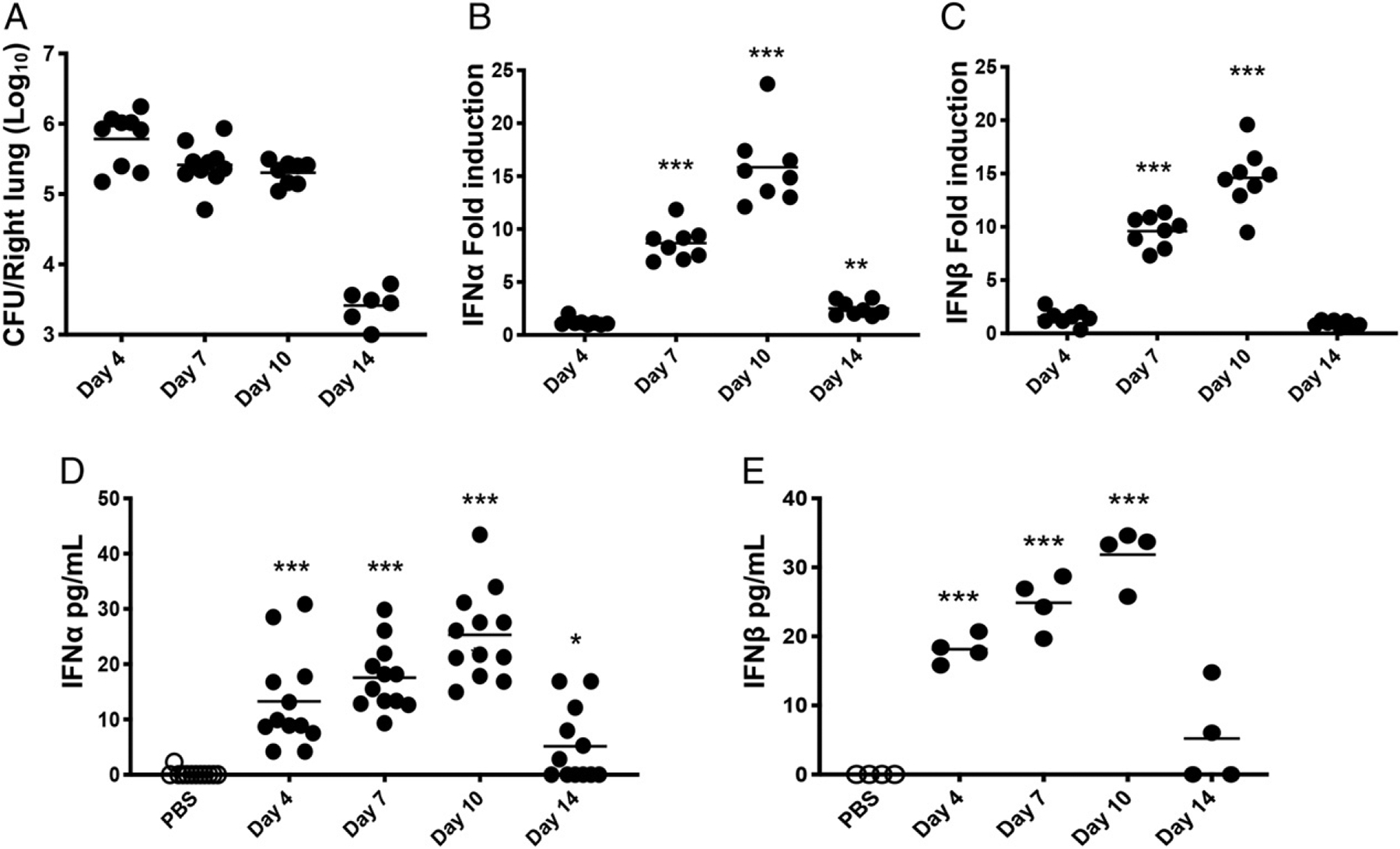
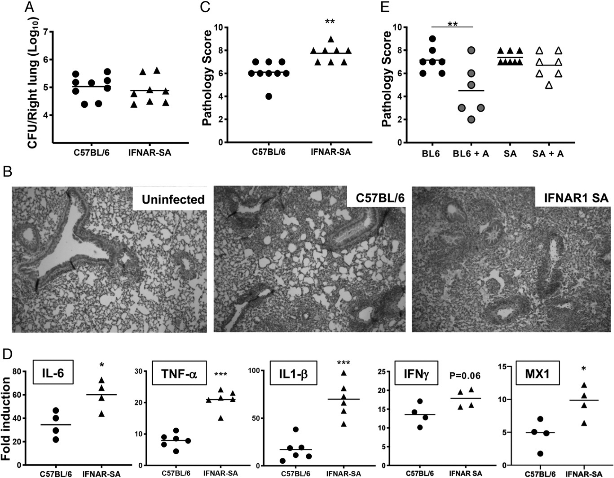
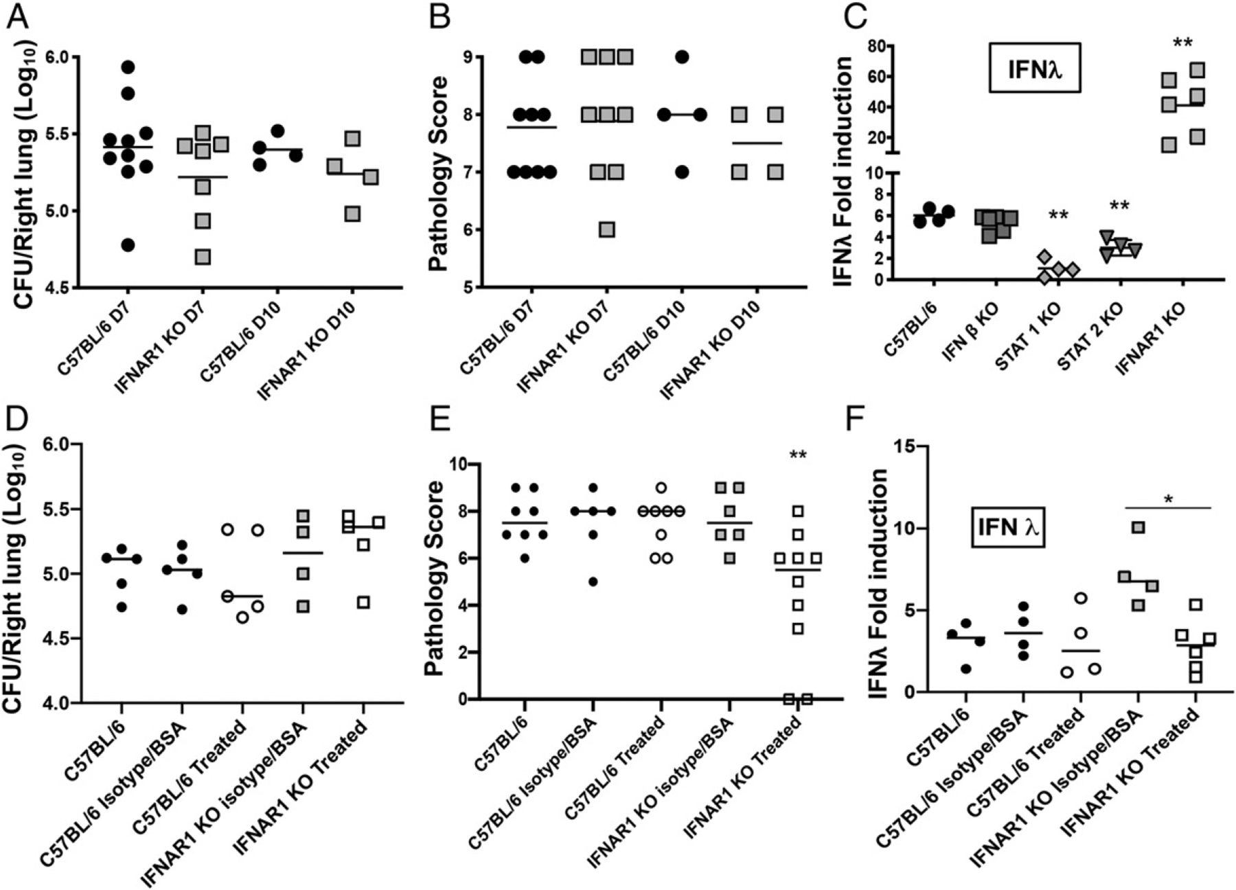
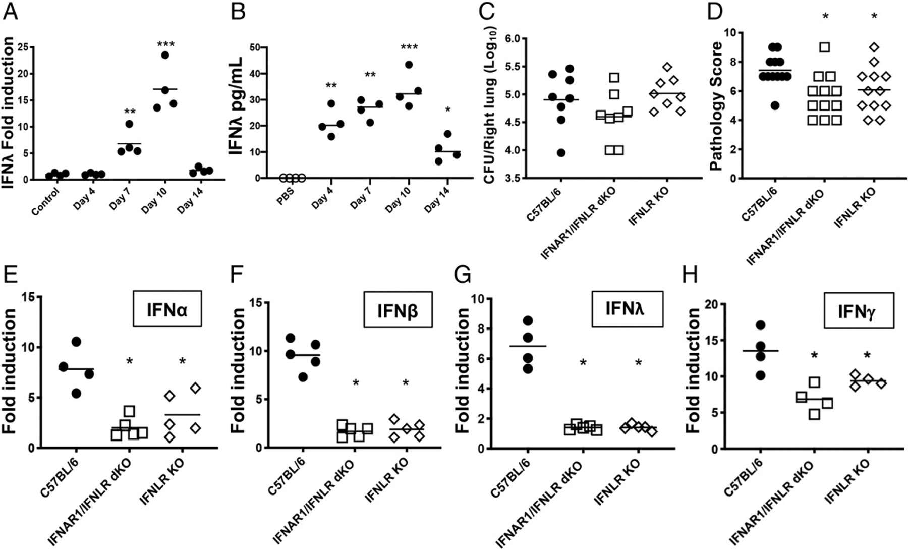
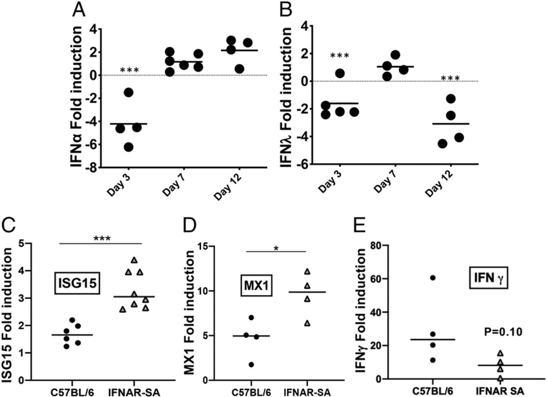
Type I and III IFNs play diverse roles in bacterial infections, being protective for some but deleterious for others. Using RNA-sequencing transcriptomics we investigated lung gene expression responses to Bordetella pertussis infection in adult mice, revealing that type I and III IFN pathways may play an important role in promoting inflammatory responses. In B. pertussis-infected mice, lung type I/III IFN responses correlated with increased proinflammatory cytokine expression and with lung inflammatory pathology. In mutant mice with increased type I IFN receptor (IFNAR) signaling, B. pertussis infection exacerbated lung inflammatory pathology, whereas knockout mice with defects in type I IFN signaling had lower levels of lung inflammation than wild-type mice. Curiously, B. pertussis-infected IFNAR1 knockout mice had wild-type levels of lung inflammatory pathology. However, in response to infection these mice had increased levels of type III IFN expression, neutralization of which reduced lung inflammation. In support of this finding, B. pertussis-infected mice with a knockout mutation in the type III IFN receptor (IFNLR1) and double IFNAR1/IFNLR1 knockout mutant mice had reduced lung inflammatory pathology compared with that in wild-type mice, indicating that type III IFN exacerbates lung inflammation. In marked contrast, infant mice did not upregulate type I or III IFNs in response to B. pertussis infection and were protected from lethal infection by increased type I IFN signaling. These results indicate age-dependent effects of type I/III IFN signaling during B. pertussis infection and suggest that these pathways represent targets for therapeutic intervention in pertussis.


Conflict of interest statement

Disclosures

The authors have no financial conflicts of interest.

Figures

FIGURE 1.
FIGURE 1.
Lung transcriptional response to B. pertussis infection in adult C57BL/6 mice. Mice (n = 4 per group) were euthanized on day 4 postinoculation with B. pertussis or PBS sham inoculum, and RNA was isolated from the lungs. RNA-seq and IPA revealed (A) total number and direction of differentially expressed genes, (B) top 10 most upregulated genes, (C) top canonical pathways represented by the differentially expressed genes, and (D) predicted upstream regulators of responses, ranked by significance (left) or z-score (right).
FIGURE 2.
FIGURE 2.
Type I IFN production induced in response to B. pertussis infection in adult mice. Mice (n ≥ 4 per group) were euthanized on the indicated dpi (x-axis) with B. pertussis or PBS sham inoculum, and lungs were dissected for assessment of (A) bacterial burdens by plating for viable counts, (B and C) mRNA levels of IFN-α and IFN-β by quantitative RT-PCR, or (D and E) IFN-α and IFN-β protein levels by ELISA. Data are representative of at least two independent experiments. *p < 0.05, ***p < 0.001 by the Student t test and two-way ANOVA.
FIGURE 3.
FIGURE 3.
Increased IFNAR1 signaling leads to exacerbated lung inflammation in B. pertussis–infected adult mice. C57BL/6 or IFNAR-SA mice (n ≥ 4 per group) were euthanized on day 7 postinoculation with B. pertussis or PBS sham inoculum, and lungs were dissected for assessment of outcomes. (A) Bacterial burdens. (B) Representative H&E-stained inflammatory pathology images (original magnification 340) of uninfected and infected C57BL/6 and infected IFNAR1-SA mouse lung sections. (C) Inflammatory pathology scores assessed from lung histology sections. (D) Fold induction (infected versus sham inoculated) of IL-6, TNF-α, IL-1β, IFN-γ, and MX1 mRNA levels. (E) Infected C57BL/6 (BL6) or IFNAR1-SA (SA) mice (n ≥ 6 per group) were treated with AAL-R (+A) or PBS intranasally 24 h postinoculation and euthanized on day 7 postinoculation for assessment of lung inflammatory pathology scores. Data are representative of at least two independent experiments. *p < 0.05, **p < 0.01, ***p < 0.001 by the Student t test and two-way ANOVA.
FIGURE 4.
FIGURE 4.
Impaired type I IFN signaling leads to a reduction in lung inflammation in adult mice. C57BL/6, IFN-β KO, STAT1 KO, and STAT2 KO mice (n ≥ 4 per group) were euthanized on day 7 postinoculation with B. pertussis or PBS sham inoculum, and lungs were dissected for assessment of outcomes. (A) Bacterial burdens. (B) Inflammatory pathology scores assessed from lung histology sections. Data are representative of at least two independent experiments. *p < 0.05, **p < 0.01, ***p < 0.001 by two-way ANOVA.
FIGURE 5.
FIGURE 5.
IFNAR1 KO mice show no significant difference in lung inflammatory pathology from WT mice, a role for type III IFNs. C57BL/6 and IFNAR1 KO mice (n ≥ 4 per group) were euthanized on day 7 or day 10 postinoculation with B. pertussis and lungs were dissected for assessment of (A) bacterial burdens, and (B) inflammatory pathology scores assessed from lung histology sections. (C) C57BL/6, IFN-β KO, STAT1 KO, STAT2 KO, and IFNAR1 KO mice (n ≥ 4 per group) were euthanized on day 7 postinoculation with B. pertussis or PBS sham inoculum, and lungs were dissected for assessment of fold induction (infected versus sham inoculated) of IFN-λ mRNA levels. (D–F) B. pertussis–infected C57BL/6 and IFNAR1 KO mice (n ≥ 4 per group) were treated on 0, 2, and 4 dpi with a neutralizing anti–IFN-λ Ab and a viral protein, Y136, previously shown to inhibit IFN-λ signaling (treated). Control-infected mice were treated with an equivalent isotype Ab and BSA. Lung bacterial burdens (D), inflammatory pathology (E), and IFN-λ mRNA levels (F) were assayed at 7 dpi. Data are representative of at least two independent experiments. *p < 0.05, **p < 0.01, ***p < 0.001 by two-way ANOVA.
FIGURE 6.
FIGURE 6.
Type III IFN is upregulated in response to B. pertussis infection and contributes to lung inflammation in adult mice. (A and B) C57BL/6 mice (n ≥ 4 per group) were euthanized on the indicated dpi (x-axis) with B. pertussis or PBS sham inoculum, and lungs were dissected for assessment of (A) fold induction (infected versus sham inoculated) of IFN-λ mRNA levels and (B) IFN-λ protein levels. (C and D) C57BL/6, IFNLR1 KO, or IFNAR1/IFNLR1 double KO mice were euthanized on day 7 postinoculation with B. pertussis, and lungs were dissected for assessment of (C) bacterial burdens and (D) inflammatory pathology. (E–H) Fold induction (infected versus sham inoculated) of mRNA levels of the indicated IFN genes at day 7 postinoculation with B. pertussis or PBS sham inoculum in adult C57BL/6, IFNLR1 KO, or IFNAR1/IFNLR1 double KO mice (n ≥ 4 per group). Data are representative of at least two independent experiments. *p < 0.05, **p < 0.01, ***p < 0.001 by Student t test and two-way ANOVA.
FIGURE 7.
FIGURE 7.
Type I and III IFNs are not upregulated by B. pertussis infection in infant WT mice. (A and B) Infant C57BL/6 mice (n ≥ 4 per group) were euthanized on the indicated dpi (x-axis) with B. pertussis or PBS sham inoculum, and lungs were dissected for assessment of fold induction (infected versus sham inoculated) of (A) IFN-α and (B) IFN-λ mRNA levels. (C–E) Infant C57BL/6 and IFNAR1-SA mice (n ≥ 6 per group) were euthanized on day 7 postinoculation with B. pertussis or PBS sham inoculum, and lungs were dissected for assessment of fold induction (infected versus sham inoculated) of the ISGs (C) ISG15 and (D) MX1 or (E) IFN-g mRNA levels. Data are representative of at least two independent experiments. *p < 0.05, **p < 0.01, ***p < 0.001 by Student t test.
FIGURE 8.
FIGURE 8.
Increased IFNAR signaling is protective against lethal B. pertussis infection in infant mice. (A) Time course of survival of infant C57BL/6 and IFNAR1-SA mice after inoculation with B. pertussis. (B) Increase in WBC counts in blood of infant C57BL/6 and IFNAR1-SA mice 7 dpi with B. pertussis. Data are representative of at least two independent experiments. **p < 0.01, Student t test or log-rank (Mantel–Cox) test (for survival curves).

References

    1. Clark TA 2014. Changing pertussis epidemiology: everything old is new again. J. Infect. Dis 209: 978–981. - PubMed
    1. Heininger U, Klich K, Stehr K, and Cherry JD. 1997. Clinical findings in Bordetella pertussis infections: results of a prospective multicenter surveillance study. Pediatrics 100: E10. - PubMed
    1. Mattoo S, and Cherry JD. 2005. Molecular pathogenesis, epidemiology, and clinical manifestations of respiratory infections due to Bordetella pertussis and other Bordetella subspecies. Clin. Microbiol. Rev 18: 326–382. - PMC - PubMed
    1. Paddock CD, Sanden GN, Cherry JD, Gal AA, Langston C, Tatti KM, Wu KH, Goldsmith CS, Greer PW, Montague JL, et al. 2008. Pathology and pathogenesis of fatal Bordetella pertussis infection in infants. Clin. Infect. Dis 47: 328–338. - PubMed
    1. Somerville RL, Grant CC, Grimwood K, Murdoch D, Graham D, Jackson P, Meates-Dennis M, Nicholson R, and Purvis D. 2007. Infants hospitalised with pertussis: estimating the true disease burden. J. Paediatr. Child Health 43: 617–622. - PubMed

Publication types

MeSH terms