. 2020 Apr 21;64(5):e00106-20.
doi: 10.1128/AAC.00106-20. Print 2020 Apr 21.

Structural Insights into Ceftobiprole Inhibition of Pseudomonas aeruginosa Penicillin-Binding Protein 3

Affiliations

Structural Insights into Ceftobiprole Inhibition of Pseudomonas aeruginosa Penicillin-Binding Protein 3

Vijay Kumar et al. Antimicrob Agents Chemother. .

Abstract

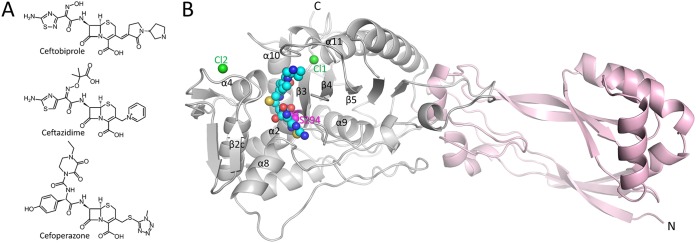
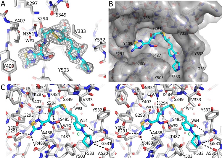
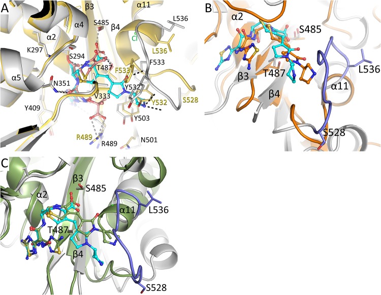
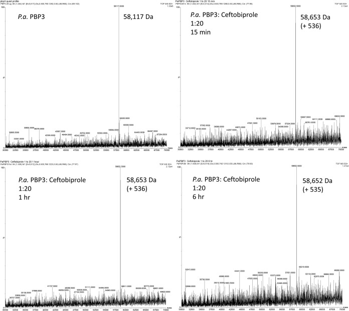
Ceftobiprole is an advanced-generation broad-spectrum cephalosporin antibiotic with potent and rapid bactericidal activity against Gram-positive pathogens, including methicillin-resistant Staphylococcus aureus, as well as susceptible Gram-negative pathogens, including Pseudomonas sp. pathogens. In the case of Pseudomonas aeruginosa, ceftobiprole acts by inhibiting P. aeruginosa penicillin-binding protein 3 (PBP3). Structural studies were pursued to elucidate the molecular details of this PBP inhibition. The crystal structure of the His-tagged PBP3-ceftobiprole complex revealed a covalent bond between the ligand and the catalytic residue S294. Ceftobiprole binding leads to large active site changes near binding sites for the pyrrolidinone and pyrrolidine rings. The S528 to L536 region adopts a conformation previously not observed in PBP3, including partial unwinding of the α11 helix. These molecular insights can lead to a deeper understanding of β-lactam-PBP interactions that result in major changes in protein structure, as well as suggesting how to fine-tune current inhibitors and to develop novel inhibitors of this PBP.

Keywords: beta-lactam antibiotic; penicillin-binding protein; protein crystallography.


Figures

FIG 1
FIG 1
Structures of ceftobiprole and PBP3. (A) Chemical structures of ceftobiprole, ceftazidime, and cefoperazone. (B) Overall structure of PaPBP3 in complex with ceftobiprole. The C-terminal catalytic transpeptidase domain (gray) and the N-terminal domain (pink) are drawn as a ribbon diagram. Atoms in ceftobiprole and the chloride ions are depicted as spheres (cyan carbons, blue nitrogens, red oxygens, yellow sulfur, and green chlorides). The catalytic S294 is shown as pink spheres and is labeled. Key secondary structural elements are labeled as in reference .
FIG 2
FIG 2
Ceftobiprole binding to PBP3. (A) Unbiased omit |Fo|-|Fc| difference density contoured at 3σ, showing the presence of the covalently bound ceftobiprole in the active site. Ceftobiprole is shown in stick representation, with cyan carbon atoms. (B) Surface representation of the PBP3 active site, with ceftobiprole bound. (C) Side-by-side stereo figure showing the interactions between ceftobiprole and the active site residues. Hydrogen bonds involving ceftobiprole atoms are depicted as dashed lines. Ceftobiprole and bound water molecules are shown in stick and spherical representation, respectively. The chloride ion (Cl-1) and water molecules (W#1 to W#4) are shown.
FIG 3
FIG 3
Structural comparison of the ceftobiprole-PBP3 structure. (A) Superposition of the ceftobiprole- and ceftazidime-bound PBP3 structures. The root mean square deviation (RMSD) of the 466 aligned C-α atoms is 1.00 Å (the ceftazidime-PaPBP3 structure has PDB accession number 3PBO [4]). The PBP3 protein residues from the ceftobiprole and ceftazidime complexes are colored gray and yellow, respectively. The cephalosporin ligands are both shown in ball-and-stick representation, ceftobiprole with cyan carbon atoms and ceftazidime with orange carbon atoms. Hydrogen bonds of ceftobiprole and ceftazidime with the protein are shown as black and gray dashed lines, respectively. Residues that differ significantly between the two structures have the two different conformations labeled in black or yellow; other residues have a single black label. Key secondary structural elements are also labeled. (B) Superposition of the ceftobiprole-bound PaPBP3 and ceftobiprole-bound Staphylococcus aureus PBP4 (PDB accession number 5TXI [14]) structures. The PBP4 protein is colored orange, as are the carbon atoms of its bound ceftobiprole; the PBP3-ceftobiprole atoms are colored as in panel A except for residues 528 to 536, which are colored light blue. The following residues were used for superpositioning of the active sites: PBP3 residues 290 to 307, 345 to 360, 406 to 409, 484 to 488, and 505 to 511 were superimposed on PBP4 residues 71 to 88, 135 to 150, 179 to 182, 259 to 263, and 268 to 274, respectively; the RMSD for 50 C-α atoms was 1.34 Å. (C) Superposition of the ceftobiprole-bound PaPBP3 and ceftobiprole-bound Staphylococcus aureus PBP2a (PDB accession number 4DKI [15]) structures. The PBP2a protein is colored green, as are the carbon atoms of its bound ceftobiprole; the PBP3-ceftobiprole atoms are colored as in panel B. The following residues were used for superpositioning of the active sites: PBP3 residues 290 to 307, 340 to 360, 406 to 409, 482 to 488, and 503 to 510 were superimposed on PBP2a residues 399 to 416, 453 to 473, 518 to 521, 595 to 601, and 613 to 620, respectively; the RMSD for 58 C-α atoms was 1.06 Å.
FIG 4
FIG 4
DSF thermal shift assay of ceftobiprole and ceftazidime binding to PaPBP3. The derivative of the change in fluorescence is plotted versus temperature. Experiments were performed in duplicate.
FIG 5
FIG 5
Mass spectra for PaPBP3 alone and after 15 min, 1 h, and 6 h of preincubation with ceftobiprole. The PaPBP3/ceftobiprole molar ratio was 1:20. Mass accuracy is ±5 Da.

References

    1. Tacconelli E, Magrini N, Carmeli Y, Harbath S, Kahlmeter G, Kluytmans J, Mendelson M, Pulcini C, Singh N, Theuretzbacher U. 2017. Global priority list of antibiotic-resistant bacteria to guide research, discovery, and development of new antibiotics. World Health Organization, Geneva, Switzerland: https://www.who.int/medicines/publications/WHO-PPL-Short_Summary_25Feb-E....
    1. Pang Z, Raudonis R, Glick BR, Lin TJ, Cheng Z. 2019. Antibiotic resistance in Pseudomonas aeruginosa: mechanisms and alternative therapeutic strategies. Biotechnol Adv 37:177–192. doi:10.1016/j.biotechadv.2018.11.013. - DOI - PubMed
    1. Chen W, Zhang YM, Davies C. 2017. Penicillin-binding protein 3 is essential for growth of Pseudomonas aeruginosa. Antimicrob Agents Chemother 61:e01651-16. doi:10.1128/AAC.01651-16. - DOI - PMC - PubMed
    1. Han S, Zaniewski RP, Marr ES, Lacey BM, Tomaras AP, Evdokimov A, Miller JR, Shanmugasundaram V. 2010. Structural basis for effectiveness of siderophore-conjugated monocarbams against clinically relevant strains of Pseudomonas aeruginosa. Proc Natl Acad Sci U S A 107:22002–22007. doi:10.1073/pnas.1013092107. - DOI - PMC - PubMed
    1. Pfaller MA, Flamm RK, Mendes RE, Streit JM, Smart JI, Hamed KA, Duncan LR, Sader HS. 2018. Ceftobiprole activity against Gram-positive and -negative pathogens collected from the United States in 2006 and 2016. Antimicrob Agents Chemother 63:e01566-18. doi:10.1128/AAC.01566-18. - DOI - PMC - PubMed

Publication types

MeSH terms