. 2020 Mar 24;117(12):6484-6490.
doi: 10.1073/pnas.1918936117. Epub 2020 Mar 9.

Visualizing the protons in a metalloenzyme electron proton transfer pathway

Affiliations

Visualizing the protons in a metalloenzyme electron proton transfer pathway

Hanna Kwon et al. Proc Natl Acad Sci U S A. .

Abstract

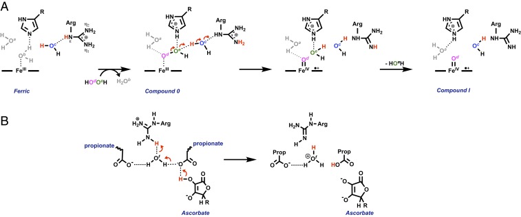
In redox metalloenzymes, the process of electron transfer often involves the concerted movement of a proton. These processes are referred to as proton-coupled electron transfer, and they underpin a wide variety of biological processes, including respiration, energy conversion, photosynthesis, and metalloenzyme catalysis. The mechanisms of proton delivery are incompletely understood, in part due to an absence of information on exact proton locations and hydrogen bonding structures in a bona fide metalloenzyme proton pathway. Here, we present a 2.1-Å neutron crystal structure of the complex formed between a redox metalloenzyme (ascorbate peroxidase) and its reducing substrate (ascorbate). In the neutron structure of the complex, the protonation states of the electron/proton donor (ascorbate) and all of the residues involved in the electron/proton transfer pathway are directly observed. This information sheds light on possible proton movements during heme-catalyzed oxygen activation, as well as on ascorbate oxidation.

Keywords: ascorbate; heme; neutron; peroxidase; proton transfer.


Conflict of interest statement

The authors declare no competing interest.

Figures

Scheme 1.
Scheme 1.
Scheme showing the chemistry of ascorbate. In the physiological pH range (shown in black), ascorbate exists as a singly deprotonated form (HA). One electron oxidation formally leads to formation of the monodehydroascorbate radical (HA), which is highly acidic [pKa = −0.45 for the OH group on C2 (52)] and releases a proton. Thus, the oxidation of ascorbate (HA) formally involves one electron and one proton (and overall one H). Other forms of ascorbate, which exist outside of the normal biological pH range, are indicated in gray.
Fig. 1.
Fig. 1.
(A) The neutron structure of the ascorbate peroxidase (APX)–ascorbate electron transfer complex. Nuclear density (2Fo − Fc) of Arg38, His42, Arg172, and ascorbate is shown in cyan (contoured at 1.5σ). For simplicity, the heme is shown in pink and at partial transparency with the hydrogen atoms shown in magenta, oxygen atoms in red, nitrogen atoms in blue, deuterium in white, and carbon in green. Arg38 is in the neutral (guanidine) form. Water molecules are indicated as W; waters b (Wb) and c (Wc) that are included in the mechanism in Fig. 3 are labeled accordingly. (B) Skeleton outline of the proton transfer pathway, highlighting only the individual deuterium atoms and hydrogen bonds involved. (C) The hydrogen bonding arrangements in the region of the heme 6-propionate and 7-propionates (labeled 6 and 7, respectively) and His169. (D) The local environment of the Arg38 residue, showing the hydrogen bonding interactions between the guanidine group and Asn72 and water molecules that are within hydrogen bonding distance of Arg38. A further water molecule (Wf) is identified (shown also Figs. 3 and 4). The δ- and γ-heme edges are labeled for reference in C and D. Hydrogen bond distances (in angstroms) are shown in gray in BD. Because of the rotational freedom of the water molecules, only the distances between hydrogen bond donor and acceptor are shown.
Fig. 2.
Fig. 2.
Nuclear density (2Fo − Fc), shown in cyan and contoured at 1.5σ for Arg38 in (A) the ferric APX–ascorbate complex and (B) ferric APX structure.
Fig. 3.
Fig. 3.
Exemplars of possible movements of protons involving Arg38, based on an analysis of neutron structures for ferric APX and the ferric APX–ascorbate complex and on the neutron structure of compound II of APX (53). In A is envisaged a series of proton transfers that occur during O–O bond cleavage and formation of compound I. Note that the distal Arg and the water molecule labeled c (in blue) swap positions between APX (shown here) and CcP (12) (the latter having Arg48 in the position of water c), which may open up alternative proton delivery pathways (see text). In B is envisaged proton delivery from the H donor (ascorbate) to Arg38 during reduction of compounds I or II. Since tautomeric forms of Arg will rapidly interconvert in solution, other combinations of proton movements are possible; this could include using neutral arginine as a catalytic base, to act as the “pull” for the concerted movement of protons from one place to another during different parts of a catalytic cycle [as previously suggested (29)]. Note also that His42 is unprotonated in both ferric APX structures (SI Appendix, Fig. S1); this is analogous to the equivalent His52 residue in CcP (12), which is also neutral. Proton delivery to the distal histidine is presumed in the mechanism here, as both His42 in APX (53) and His52 in CcP (12) are protonated after reaction with H2O2. Individual water molecules are labeled as af and colored accordingly; the structural positions of water molecules ac and f are indicated in Fig. 1 and SI Appendix, Fig. S1. The proposed structure shown for compound 0 is the same as that identified by computation (54) and the proposed hydrogen bond identified computationally shown in gray.
Fig. 4.
Fig. 4.
Comparison of possible proton channels. (A) The figure shows a channel of water molecules extending out from the heme in the direction of the δ-heme edge in the ferric APX–ascorbate complex, which may function as an alternative proton delivery pathway. The waters are positioned within hydrogen-bonding range of one another (∼2.8 Å), with the exception of the two outer waters (labeled *), which are ∼3.8 Å from their adjacent waters. The distance between the iron of the heme group and the two outer waters is ∼12.4 Å. Our expectation is that water molecules move slightly and are not static in solution, as is evidenced by the fact that the water positions in the ferric APX–ascorbate complex (shown in red for oxygen and white for deuterium) are slightly altered from those in the ferric-only structure (overlaid in cyan/white). Compare also the slightly different orientations of conserved water molecules (SI Appendix, Fig. S1 A and B). The color scheme is the same as that shown in Fig. 1. (B) For comparison, the schematic shows proposed proton transfer pathways (H, K, and D channels) in bovine heart cytochrome c oxidase [PDB ID code 5B1A (55)]. The D channel connects D91 to E242; the K channel connects to Y244; and the H channel connects to D51 via the pyrrole of heme (redrawn from ref. 56). Water molecules identified in the crystal structure are shown as red spheres.

References

    1. Warren J. J., Tronic T. A., Mayer J. M., Thermochemistry of proton-coupled electron transfer reagents and its implications. Chem. Rev. 110, 6961–7001 (2010). - PMC - PubMed
    1. Parada G. A., et al. , Concerted proton-electron transfer reactions in the Marcus inverted region. Science 364, 471–475 (2019). - PMC - PubMed
    1. Tsukihara T., et al. , The whole structure of the 13-subunit oxidized cytochrome c oxidase at 2.8 Å. Science 272, 1136–1144 (1996). - PubMed
    1. Iwata S., Ostermeier C., Ludwig B., Michel H., Structure at 2.8 Å resolution of cytochrome c oxidase from Paracoccus denitrificans. Nature 376, 660–669 (1995). - PubMed
    1. Buschmann S., et al. , The structure of cbb3 cytochrome oxidase provides insights into proton pumping. Science 329, 327–330 (2010). - PubMed

Publication types

LinkOut - more resources