. 2020 Apr 20;88(5):e00841-19.
doi: 10.1128/IAI.00841-19. Print 2020 Apr 20.

Fluorescence-Reported Allelic Exchange Mutagenesis-Mediated Gene Deletion Indicates a Requirement for Chlamydia trachomatis Tarp during In Vivo Infectivity and Reveals a Specific Role for the C Terminus during Cellular Invasion

Affiliations

Fluorescence-Reported Allelic Exchange Mutagenesis-Mediated Gene Deletion Indicates a Requirement for Chlamydia trachomatis Tarp during In Vivo Infectivity and Reveals a Specific Role for the C Terminus during Cellular Invasion

Susmita Ghosh et al. Infect Immun. .

Abstract

The translocated actin recruiting phosphoprotein (Tarp) is a multidomain type III secreted effector used by Chlamydia trachomatis In aggregate, existing data suggest a role of this effector in initiating new infections. As new genetic tools began to emerge to study chlamydial genes in vivo, we speculated as to what degree Tarp function contributes to Chlamydia's ability to parasitize mammalian host cells. To address this question, we generated a complete tarP deletion mutant using the fluorescence-reported allelic exchange mutagenesis (FRAEM) technique and complemented the mutant in trans with wild-type tarP or mutant tarP alleles engineered to harbor in-frame domain deletions. We provide evidence for the significant role of Tarp in C. trachomatis invasion of host cells. Complementation studies indicate that the C-terminal filamentous actin (F-actin)-binding domains are responsible for Tarp-mediated invasion efficiency. Wild-type C. trachomatis entry into HeLa cells resulted in host cell shape changes, whereas the tarP mutant did not. Finally, using a novel cis complementation approach, C. trachomatis lacking tarP demonstrated significant attenuation in a murine genital tract infection model. Together, these data provide definitive genetic evidence for the critical role of the Tarp F-actin-binding domains in host cell invasion and for the Tarp effector as a bona fide C. trachomatis virulence factor.

Keywords: Chlamydia trachomatis; Tarp; actin; actin cytoskeleton; cytoskeleton; effector; effector functions.


Figures

FIG 1
FIG 1
Generation of C. trachomatis ΔtarP. (A) Schematic of the tarP locus in C. trachomatis L2. FRAEM was used to delete the entire tarP (ctl0716) sequence and insert a cassette containing gfp and bla. (B) The loss of tarP and presence of gfp were confirmed in the resulting strain via qPCR. Curing of pSUmC was confirmed using primers specific for mcherry, and retention of endogenous pL2 was verified using primers specific for plasmid-encoded pgp7-8. (C) Chemiluminescence immunoblot of EB material from wild-type (WT) or mutant Chlamydia (tarp) with TarP-specific antibodies. MOMP was probed as a loading control. Cultures of HeLa cells were infected with equal IFU of WT or tarP chlamydiae. At 0, 12, 24, 36, and 48 h postinfection, one replicate was processed for enumeration of progeny EBs on fresh HeLa monolayers (D) while the other was fixed and stained for measurement of inclusion areas (24 h is shown) (E).
FIG 2
FIG 2
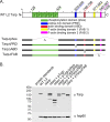
Schematic representation of Tarp and the complementation clones expressed in the tarP mutant. (A) Chlamydia trachomatis (L2) Tarp harbors an N-terminal tyrosine-rich repeat region, which is also referred to as the phosphorylation domain (phos; YYY, green boxes), a proline-rich domain (PRD; blue box), a single G-actin-binding domain (ABD; red box), and two F-actin-binding domains (yellow box [FAB 1] and pink box [FAB 2]). In-frame deletions engineered to remove specific protein domains for complementation are indicated with hatch marks and the corresponding Δ designation (Δphos, ΔPRD, ΔABD, ΔFAB 1, and ΔFAB 2). Numbers along the top of the schematic indicate amino acid positions encoded within the C. trachomatis L2 tarP gene. (B) Protein extracts from density gradient purified wild-type C. trachomatis L2 (WT L2), tarP deletion mutant (Δtarp), and mutants complemented from plasmid-based expression employing the Tarp promoter with full-length tarPtarp + pTarp), tarP lacking the phosphorylation domain (Δtarp + pTarpΔphos), tarP lacking the proline-rich domain (Δtarp + pTarpΔPRD), tarP lacking the actin-binding domain (Δtarp + pTarpΔABD), or tarP lacking the F-actin-binding domains (Δtarp + pTarpΔFAB). EBs were resolved by SDS-PAGE and visualized by immunoblotting analysis with Tarp (α Tarp) and C. trachomatis heat shock protein 60 (α Hsp60)-specific antibodies. The molecular masses of protein standards are shown.
FIG 3
FIG 3
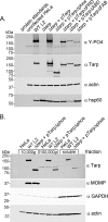
Tarp secretion and phosphorylation by Chlamydia trachomatis L2 tarP mutant and complemented clones. (A) Protein lysates were generated from HeLa cells infected with wild-type C. trachomatis L2 (WT L2), a tarP deletion mutant (Δtarp), and mutants complemented with full-length tarPtarp + pTarp), tarP lacking the phosphorylation domain (Δtarp + pTarpΔphos), tarP lacking the proline-rich domain (Δtarp + pTarpΔPRD), tarP lacking the actin-binding domain (Δtarp + pTarpΔABD), or tarP lacking the F-actin-binding domains (Δtarp + pTarpΔFAB). Protein samples underwent immunoblotting analysis with phosphotyrosine (α Y-PO4), Tarp (α Tarp), actin (α actin), and C. trachomatis heat shock protein 60 (α Hsp60)-specific antibodies. A protein lysate generated from uninfected HeLa cells (uninfected HeLa) served as a negative control. (B) Subcellular fractionation of C. trachomatis-infected cells by differential centrifugation out to 100,000 × g yields a soluble Tarp fraction that is distinct from intact elementary bodies. Total lysates derived from HeLa cells alone or HeLa cells infected with wild-type C. trachomatis L2 (WT L2) or the tarP mutant complemented with pTarpΔphos (Δtarp + pTarpΔphos) underwent subcellular fractionation by centrifugation. Fractions were resolved by SDS-PAGE and transferred to nitrocellulose for immunoblotting analysis with antibodies specific for phosphotyrosine (α Y-PO4); Tarp (α Tarp); C. trachomatis major outer membrane protein (α MOMP); glyceraldehyde-3-phosphate dehydrogenase, a soluble protein marker (α GAPDH); and actin, a protein expected to be present in all fractions (α actin).
FIG 4
FIG 4
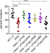
The C. trachomatis tarP mutant is deficient in chlamydial entry (and the C-terminal F-actin-binding sites play a key role in invasion). Wild-type Chlamydia trachomatis (WT L2, black circles), C. trachomatis tarP mutant (Δtarp, red squares), and C. trachomatis tarP mutant complemented with pTarp (Δtarp + pTarp, green triangles), pTarpΔphos (Δtarp +pTarpΔphos, blue circles), pTarpΔPRD (Δtarp + pTarpΔPRD, yellow diamonds), pTarpΔABD (Δtarp + pTarpΔABD, purple circles), or pTarpΔFAB (Δtarp + pTarpΔFAB, black squares) were examined for chlamydial invasion of HeLa 229 cells after 1 h. The graph presented is from one representative experiment of three. Data sets were compared with one-way ANOVA and Tukey’s multiple comparison test of the mean. *, P < 0.05; ****, P < 0.0001.
FIG 5
FIG 5
Chlamydia trachomatis invasion of HeLa cells alters the host cell shape. (A) Wild-type Chlamydia trachomatis L2 (WT L2, light green squares), tarP mutant (ΔtarP, red triangles), or tarP mutant complemented with pTarp (Δtarp + pTarp, blue upside down triangles) underwent chlamydial invasion of HeLa 229 cells for 30 min. Bacteria were visualized by staining with anti-MOMP primary antibody followed by goat anti-mouse Alexa Fluor 488 secondary antibody. The actin cytoskeleton was stained with Alexa Fluor 568 Phalloidin. The cell thickness was compared to uninfected HeLa cells (uninfected HeLa, green circles) by z-stack analysis on a Zeiss 710 inverted confocal microscope. Samples were compared using one-way ANOVA and Tukey’s multiple comparison test. ****, P < 0.0001. The graph presented is from one representative experiment of three performed. Host cells pretreated with the actin-destabilizing drug cytochalasin D (uninfected HeLa + cyto D, black circles) or drug-treated cells infected with wild-type C. trachomatis (WT L2 + cyto D, black triangles) did not undergo changes to cell shape. (B) Similarly, HeLa 229 cells were infected with wild-type Chlamydia trachomatis L2 (WT L2, black circles), tarP mutant (Δtarp, black squares), or tarP mutant complemented with pTarpΔphos (Δtarp + pTarpΔphos, black triangles) or pTarpΔFAB (Δtarp + pTarpΔFAB, black diamonds), and the cell thickness was quantified as described in panel A.
FIG 6
FIG 6
Attenuation of C. trachomatis tarP in a murine model can be reversed by cis complementation with WT tarP. (A) Schematic of allelic replacement strategy to restore tarP to the mutant strain. FRAEM was used to replace the gfp-bla cassette in the ΔtarP mutant with the tarP gene and a downstream aadA selection marker. (B) Representative fluorescence-based Western blot of tarP levels in 24-h culture material derived from equal infections with wild-type (WT), mutant (tarp), and cis-complemented (cis-tarp) Chlamydia. Hsp60 was probed as a loading control. (C) Signal intensity of Tarp-specific signal was normalized to Hsp60, and values are plotted as a function WT Tarp signal. Error bars represent one standard deviation from blots derived from 3 separate experiments. Groups of 5 female C3H/HeJ mice were infected intravaginally with equal input IFUs, and shed IFUs were enumerated every 4 days from day 3 to 31 postinfection. Shed bacteria were enumerated by passage in HeLa cells and are represented as number of mice actively shedding detectible chlamydiae (D) or numbers of detected inclusions (E). Data are represented with standard deviation, and two-way repeated measures (RM) ANOVA was performed to establish statistical significance (*, P < 0.0001) compared to the wild type.

Similar articles

Cited by

References

    1. Moulder JW, Hatch TP, Kuo CC, Schachter J, Storz J. 1984. Chlamydia, p 729–739. In Krieg NR. (ed), Bergey’s manual of systematic bacteriology. Williams & Wilkins, Baltimore, MD.
    1. Pizarro-Cerdá J, Cossart P. 2006. Bacterial adhesion and entry into host cells. Cell 124:715–727. doi:10.1016/j.cell.2006.02.012. - DOI - PubMed
    1. Mueller KE, Plano GV, Fields KA. 2014. New frontiers in type III secretion biology: the Chlamydia perspective. Infect Immun 82:2–9. doi:10.1128/IAI.00917-13. - DOI - PMC - PubMed
    1. Ferrell JC, Fields KA. 2016. A working model for the type III secretion mechanism in Chlamydia. Microbes Infect 18:84–92. doi:10.1016/j.micinf.2015.10.006. - DOI - PMC - PubMed
    1. Clifton DR, Fields KA, Grieshaber SS, Dooley CA, Fischer ER, Mead DJ, Carabeo RA, Hackstadt T. 2004. A chlamydial type III translocated protein is tyrosine-phosphorylated at the site of entry and associated with recruitment of actin. Proc Natl Acad Sci U S A 101:10166–10171. doi:10.1073/pnas.0402829101. - DOI - PMC - PubMed

Publication types