Review
. 2020 Mar 10:21:e920489.
doi: 10.12659/AJCR.920489.

Plasma Cell Myeloma with an Aggressive Clinical Course and Anaplastic Morphology in a 22-Year-Old Patient: A Case Report and Review of Literature

Affiliations
Review

Plasma Cell Myeloma with an Aggressive Clinical Course and Anaplastic Morphology in a 22-Year-Old Patient: A Case Report and Review of Literature

Hesham Elsabah et al. Am J Case Rep. .

Abstract

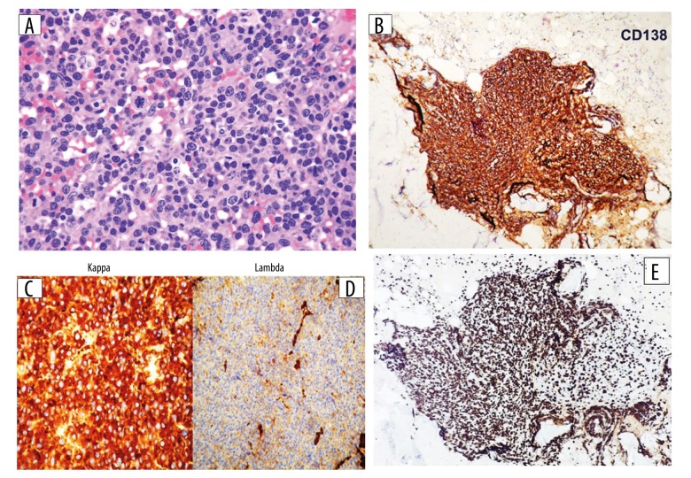
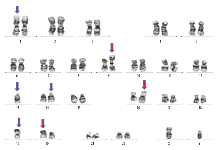
BACKGROUND Plasma cell myeloma is a neoplastic plasma cell disorder that usually presents after the fifth decade of life; it is rarely described in younger population especially under 30 years of age. However, there are conflicting reports in the literature about the clinical behavior and overall survival in younger age groups. In approximately 2% of plasma cell myeloma, the morphology of the neoplastic cells is highly pleomorphic, quite anaplastic, and may resemble metastatic tumor cells. While this poses a challenge for morphological interpretation during diagnosis, it has been demonstrated that bone marrow morphologic features (including diffuse sheet growth pattern, immature cell morphology and high mitotic index) significantly correlates with high risk disease. Moreover, there is limited description available about the morphology of the neoplastic cells when correlating the age at presentation with the clinical outcome/biological behavior; hence, the need to report and collect such cases. CASE REPORT We report a case of plasma cell myeloma in a 22-year-old male who presented with non-specific clinical features and posed a diagnostic challenge during clinical, radiological, and laboratory examination. The pathology specimens showed anaplastic morphology. Unfortunately, after diagnosis, despite treatment with brotezomib, his disease had an aggressive clinical course and he passed away 4 months after diagnosis. CONCLUSIONS Although plasma cell myeloma is rare in patients younger than 30 years, it must be considered in the differential diagnosis and investigated properly especially in patients with clinical suspicion of a metastatic non-hematological tumor. The anaplastic variant in a young patient is a diagnostic challenge and is associated with bizarre morphology, aggressive presentation, adverse cytogenetics, resistance to chemotherapy, and poor, short-term, survival.


Conflict of interest statement

Conflict of interest: None declared

Conflict of interests

None.

Figures

Figure 1.
Figure 1.
Computed tomography (CT) scan. (A) Selective axial CT image showing expanding soft tissue density mass lesion with underlying right rib osseous destruction. (B) A small lytic lesion is noted within the diploic space of the left posterior bony calvarium.
Figure 2.
Figure 2.
Magnetic resonance imaging (MRI). (A) Coronal SITR of the sacrum and sacroiliac joints showing multiple bright foci of infiltration are noted bilaterally involving the sacrum and iliac side of both sacroiliac joints. (B) Sagittal reconstruction of the dorso-lumbosacral spine (bone window setting) showing multiple hypodense lytic lesions notably at L5, D11, D10, D9 as well as the proximal sacral pieces.
Figure 3.
Figure 3.
(A) Peripheral blood smear (50×) showing few circulating neoplastic cells of blastoid morphology. (B) Bone marrow aspirate smear showing cohesive clusters/sheets of neoplastic cells mimicking metastatic tumors. (C–F) The malignant cells are large to giant in size with irregular cytoplasmic boundaries, bizarre shaped nuclei with markedly irregular contours (multilobated/abnormally lobated or multinucleated) with hyperchromatic nuclei and very prominent nucleoli (100×).(G) Some scattered plasma cells with hyperchromatic nuclei (red arrows) with frequent mitotic figures noted (black arrows).
Figure 4.
Figure 4.
Flow cytometry analysis revealed a population comprising approximately 11% of the total showing moderate side light scatter and high heterogeneous forward light scatter and negative for CD45. The population was positive for CD38 and CD138 indicative of plasma cell showing cytoplasmic kappa light chain restriction (monotypic) with aberrant surface kappa light chain expression and negative for CD56 and CD117.
Figure 5.
Figure 5.
(A) Bone marrow biopsy specimen with hematoxylin and eosin stain (20×) shows core biopsy is diffusely infiltrated by sheets of neoplastic plasma cells. The cells are markedly pleomorphic, with marked nuclear irregularities, prominent nucleoli and some mitotic figures (black arrow). (B–E) Immunohistochemistry on core biopsy specimen (10×) shows (B) CD138 positive, (C) kappa: positive, (D) lambda: negative, and (E) Ki-67 (>90%).
Figure 6.
Figure 6.
Representative karyotype of a metaphase cell with multiple numeric and structural abnormalities. ISCN nomenclature: 44,XY,i(9)(q10),-13,add(14)(q32),der(16)add(16)(p13q24),der(19)add(19)(p13.3),der(20)t(20;?)(p11;?)[20]/44,XY,dup(1) (q21q32),-13,add(14)(q32), der(16)add(16)(p13,q24),-19[6]/46,XY[4].

References

    1. Michels TC, Petersen KE. Multiple myeloma: Diagnosis and treatment. Am Fam Physician. 2017;95(6):373–83. - PubMed
    1. Kyle RA, Gertz MA, Witzig TE, et al. Review of 1027 patients with newly diagnosed multiple myeloma. Mayo Clin Proc. 2003;78(1):21–33. - PubMed
    1. Ludwig H, Durie BG, Bolejack V, et al. Myeloma in patients younger than age 50 years presents with more favorable features and shows better survival: An analysis of 10 549 patients from the International Myeloma Working Group. Blood. 2008;111(8):4039–47. - PMC - PubMed
    1. Masahiko F. The histopathology of myeloma in the bone marrow. J Clin Exp Hematop. 2018;58(2):61–67. - PMC - PubMed
    1. Rajkumar SV, Dimopoulos MA, Palumbo A, et al. International Myeloma Working Group updated criteria for the diagnosis of multiple myeloma. Lancet Oncol. 2014;15(12):e538–48. - PubMed