Review
. 2020 May;16(5):285-296.
doi: 10.1038/s41582-020-0321-y. Epub 2020 Mar 9.

Inflammation in acquired hydrocephalus: pathogenic mechanisms and therapeutic targets

Affiliations
Review

Inflammation in acquired hydrocephalus: pathogenic mechanisms and therapeutic targets

Jason K Karimy et al. Nat Rev Neurol. 2020 May.

Abstract

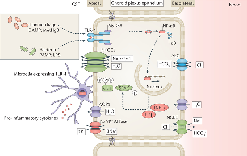
Hydrocephalus is the most common neurosurgical disorder worldwide and is characterized by enlargement of the cerebrospinal fluid (CSF)-filled brain ventricles resulting from failed CSF homeostasis. Since the 1840s, physicians have observed inflammation in the brain and the CSF spaces in both posthaemorrhagic hydrocephalus (PHH) and postinfectious hydrocephalus (PIH). Reparative inflammation is an important protective response that eliminates foreign organisms, damaged cells and physical irritants; however, inappropriately triggered or sustained inflammation can respectively initiate or propagate disease. Recent data have begun to uncover the molecular mechanisms by which inflammation - driven by Toll-like receptor 4-regulated cytokines, immune cells and signalling pathways - contributes to the pathogenesis of hydrocephalus. We propose that therapeutic approaches that target inflammatory mediators in both PHH and PIH could address the multiple drivers of disease, including choroid plexus CSF hypersecretion, ependymal denudation, and damage and scarring of intraventricular and parenchymal (glia-lymphatic) CSF pathways. Here, we review the evidence for a prominent role of inflammation in the pathogenic mechanism of PHH and PIH and highlight promising targets for therapeutic intervention. Focusing research efforts on inflammation could shift our view of hydrocephalus from that of a lifelong neurosurgical disorder to that of a preventable neuroinflammatory condition.


Conflict of interest statement

Competing interests

The authors declare no competing interests.

Figures

Figure 1 |
Figure 1 |. Classification and treatment of hydrocephalus.
Hydrocephalus can be dividied into primary and acquired forms. Hemorrhage and infection arc two of the most common causes of hydrocephalus worldwide. Both primary and acquired forms of hydrocephalus can involve intraventricular obstruction of CSF flow, which can be treated with a ventriculo-peritoneal shunt or endoscopic third ventriculostomy (ETV). ETV can be performed with or without choroid plexus coagulation (CPC). To date, all treatments for hydrocephalus are surgical, and have a high morbidity and failure rate.
Figure 2.
Figure 2.. Proposed mechanism of CSF hypersecretion in PHH and PIH
Host-derived danger-associated molecular patterns (DAMPs) such as methemoglobin (metHgB) enter the CSF during intraventricular haemorrhage and pathogen-associated molecular patterns (PAMPs), such as lipopolysaccharide (LPS) enter the CSF during bacterial meningitis. These DAMPs and PAMPs are thought to bind toll-like receptor 4 (TLR4) on the surface of the choroid plexus epithelium (CPe). This binding stimulates a TLR-4-MyD88 signalling cascade leading to nuclear translocation of nuclear factorkB (NF-κB). Nuclear NF-κB stimulates production of pro-inflammatory cytokines, for example, tumour necrosis factor-α (TNF-α) and interleukin 1β (IL-1β), which increase activity of Ste20-type stress kinase (SPAK). SPAK phosphorylates its canonical substrate, the Na+/K+/2Cl ion co-transporter- (NKCC1), and probably also phosphorylates other ion transporter targets. NKCC1 phosphorylation increases activity of the transporter, which results in a net increase in cerebrospinal fluid (CSF) production by the CPe. DAMPs and PAMPs in the CSF also bind to TLR-4 on the surface of microglial cells that are resident on the choroid plexus. This binding results in the production of pro-inflammatory cytokines by the microglia. These cytokines can bind receptors on the CPe and likely progogate CPe inflammation and CSF hypersecretion. AQP1, AE2 and NCBE are some additional transporter proteins that facilitate the passage of water (AQP1) and ions (AE2 and NCBE) across the plasma membrane. Cl
Figure 3.
Figure 3.. Glymphatic CSF transport
The glymphatic system is a perivascular cerebrospinal fliud (CSF) and interstitial fluid (ISF) exchange network that mediates waste clearance and CSF efflux from the brain to outlets such as the cervical or meningeal lymphatic system and the major draining venous sinuses. In the glymphatic system, as arteries on the surface of the cortex penetrate the brain, CSF enters the parenchyma alongside the vessels, ensheathed by astroglial endfeet. Driven by cardiac-driven arteriole pulsations, and facilitated by the high expression of aquaporin 4 (AQP4) in the astroglial endfeet, CSF exits the perivascular space and mixes with the brain’s ISF. Either by bulk flow or diffusion, the mixture of CSF and ISF flows through the parenchyma into either perivenous or perineural spaces (perineural spaces not illustrated). The fluid then travels along the perivenous or perineural spaces until it drains into to the dural sinuses or lymphatic vessels on the way to the general circulation for clearance.
Figure 4:
Figure 4:. Proposed inflammatory contributors to PHH and PIH.
Illustration showing the relative contribution of inflammation to hydrocephalus in the days week and months following haemorrhage or bacterial infection. CNS exposure to foreign pathogen-derived damage-associated molecular patterns (PAMPs), for example bacterial cell wall fragments, or host-derived damage-associated molecular patterns (DAMPs), for example blood-breakdown products, leads to an acute inflammatory response (red) in the CSF that takes place in the days to weeks after haemorrhage or infection. This response is characterized by recruitment of immune cells (e.g. microglia) and TLR4-dependent CSF hypersecretion by the CPe. Tissue damage, including friability and denudation at CSFbrain (ependyma) and CSF-blood (CPe) barrier sites, is likely to propagate and sustain the initial infectious or traumatic insult via release of other DAMPs, resulting in a transition from acute reparative inflammation (red) to chronic pathological inflammation (blue). This chronic inflammation is likely to result in scarring and obstruction of CSF drainage pathways (e.g., brain parenchymal glymphatics and meningeal lymphatics; green), which would impair CSF reabsorption. Early modulation of TLR4 activity in post-infectious and post-haemorrhagic hydrocephalus could reduce the acute CPe hypersecretory response, and prevent chronic inflammation-induced scarring. In addition, anti-inflammatory therapies offer the potential advantage of preventing the need for surgical CSF diversion, and alleviating inflammation-induced brain damage that contributes to poor long-term neurodevelopmental outcomes.

References

    1. Rekate HL A contemporary definition and classification of hydrocephalus. Seminars in pediatric neurology 16, 9–15 (2009). - PubMed
    1. Benveniste H, Lee H & Volkow ND The Glymphatic Pathway: Waste Removal from the CNS via Cerebrospinal Fluid Transport. Neuroscientist 23, 454–465 (2017). - PMC - PubMed
    1. Brinker T, Stopa E, Morrison J & Klinge P A new look at cerebrospinal fluid circulation. Fluids and barriers of the CNS 11, 10 (2014). - PMC - PubMed
    1. Furey CG, et al. De novo mutation in genes regulating neural stem cell fate in human congenital hydrocephalus. Neuron 99, 302–314e304 (2018). - PMC - PubMed
    1. Kahle KT, Kulkarni AV, Limbrick DD Jr. & Warf BC Hydrocephalus in children. Lancet 387, 788–799 (2016). - PubMed

Publication types

Substances