. 2020 Jun;16(6):653-659.
doi: 10.1038/s41589-020-0480-6. Epub 2020 Mar 9.

Deep mutational scanning reveals the structural basis for α-synuclein activity

Affiliations

Deep mutational scanning reveals the structural basis for α-synuclein activity

Robert W Newberry et al. Nat Chem Biol. 2020 Jun.

Abstract

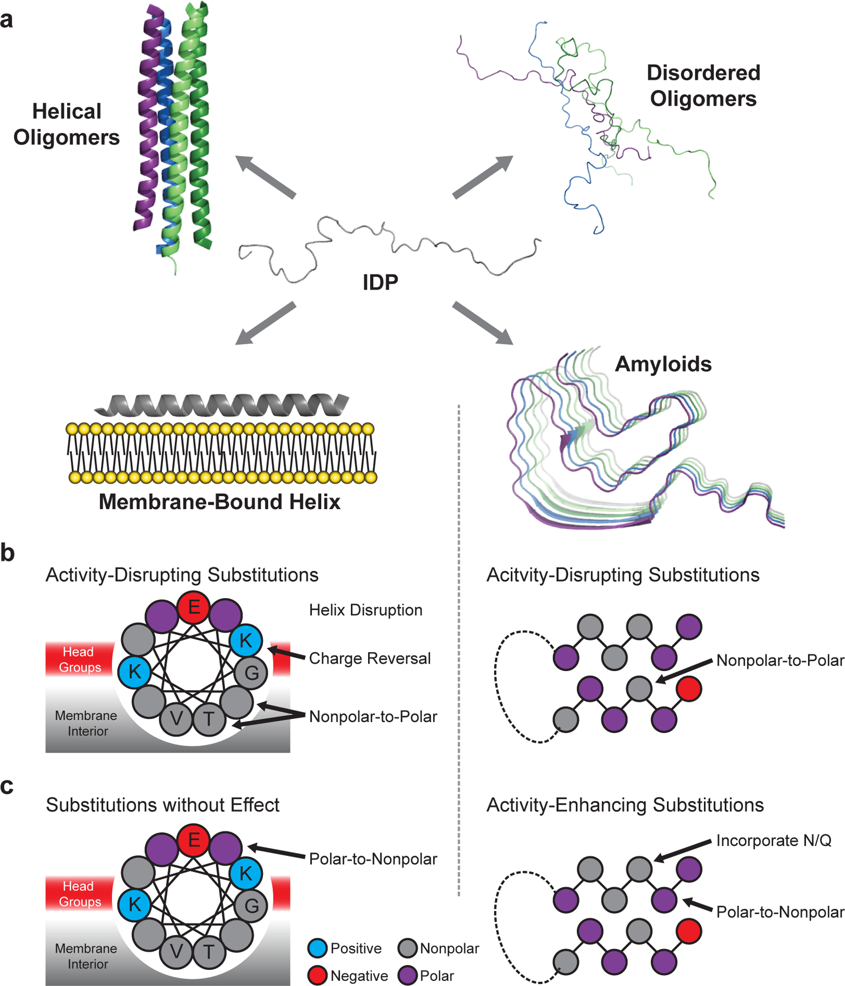
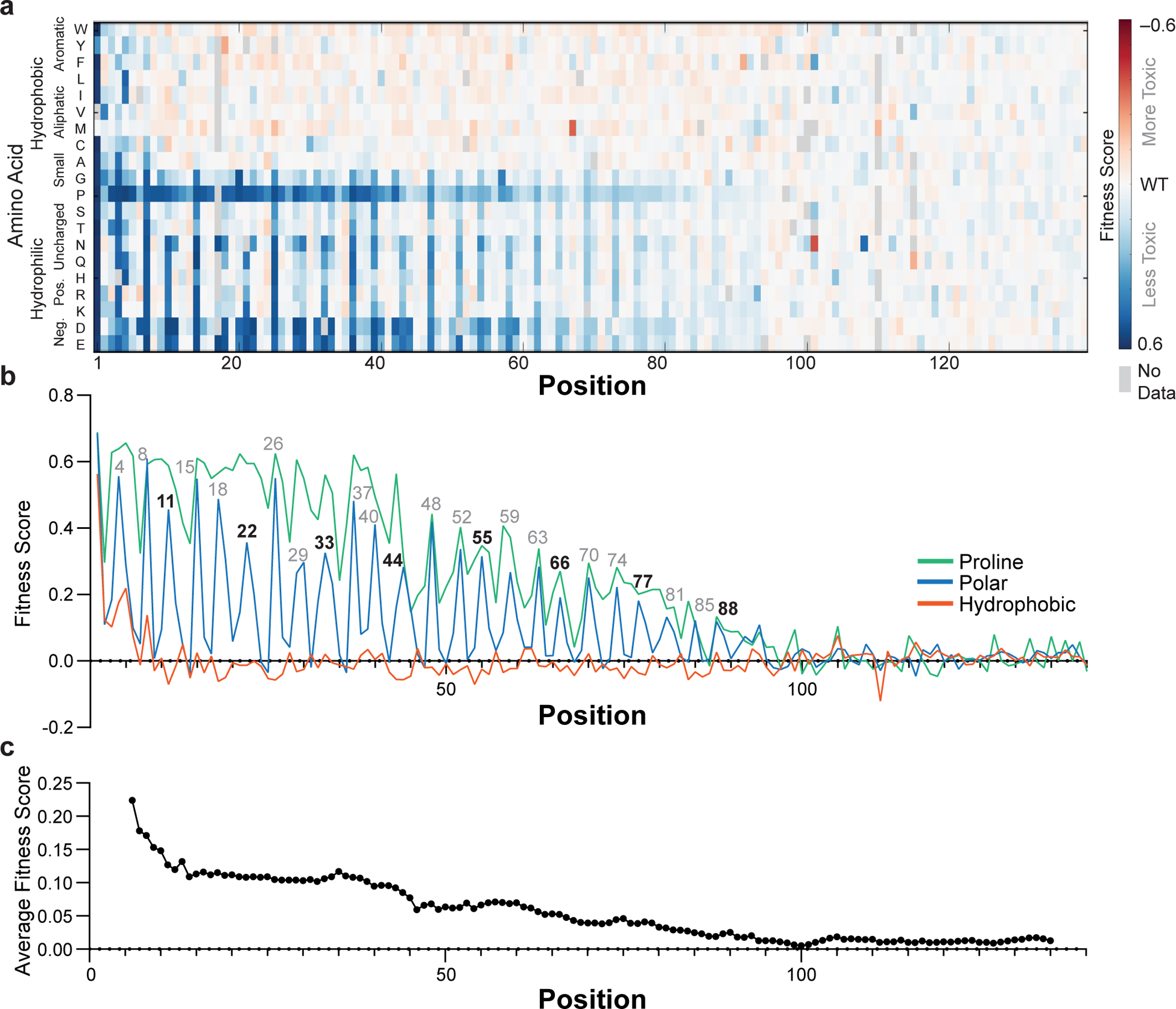
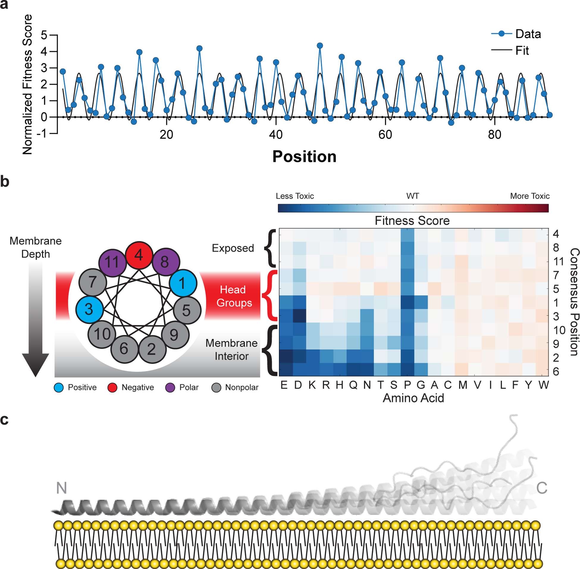
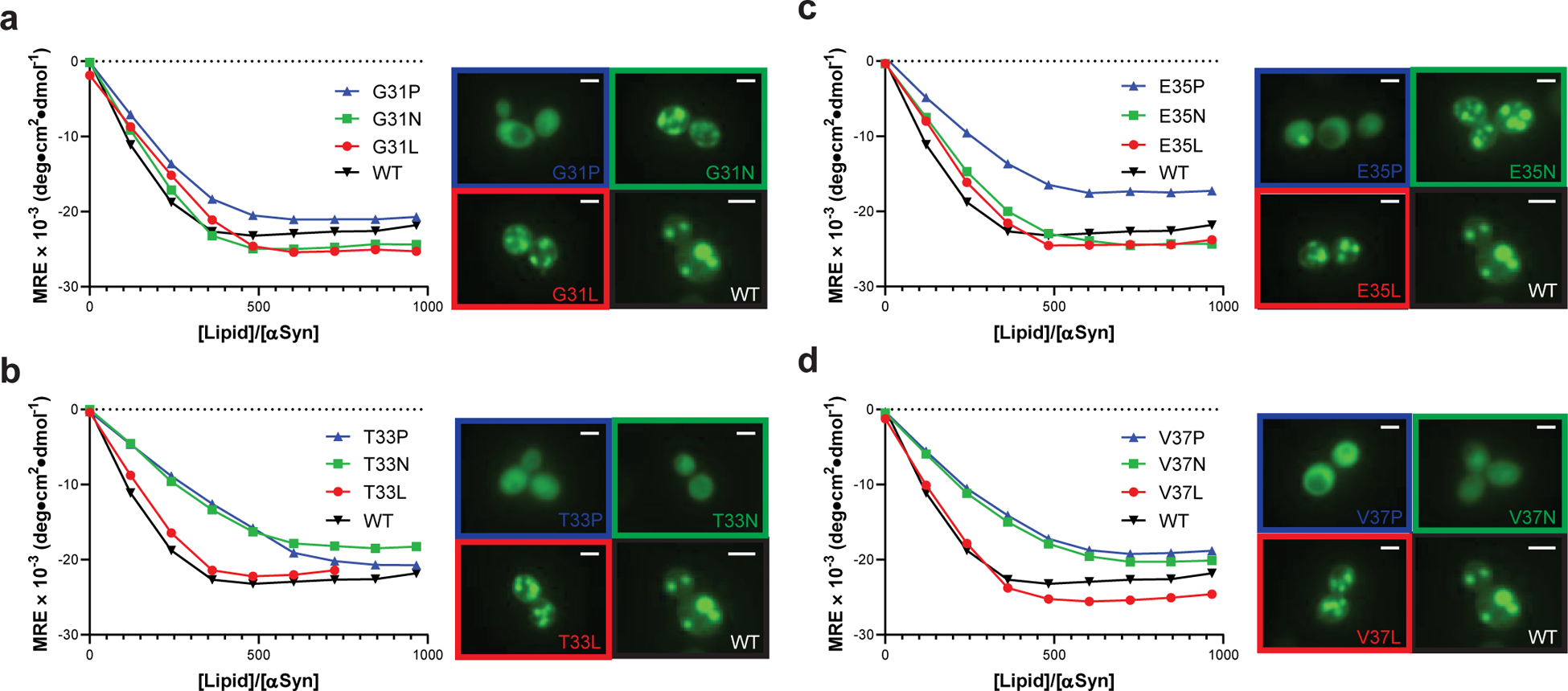
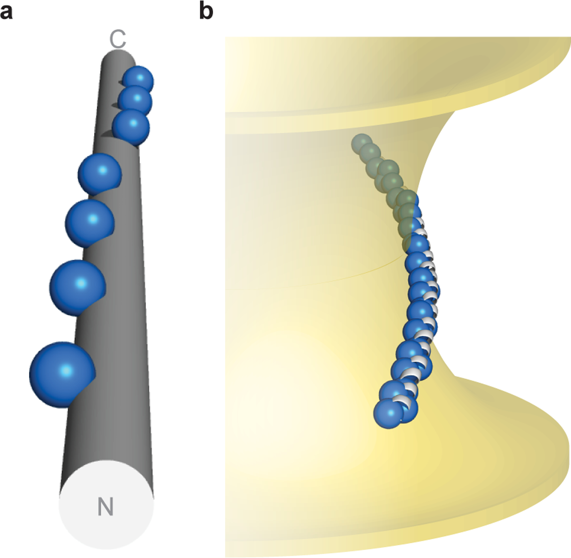
Defining the biologically active structures of proteins in their cellular environments remains challenging for proteins with multiple conformations and functions, where only a minor conformer might be associated with a given function. Here, we use deep mutational scanning to probe the structure and dynamics of α-synuclein, a protein known to adopt disordered, helical and amyloid conformations. We examined the effects of 2,600 single-residue substitutions on the ability of intracellularly expressed α-synuclein to slow the growth of yeast. Computational analysis of the data showed that the conformation responsible for this phenotype is a long, uninterrupted, amphiphilic helix with increasing dynamics toward the C terminus. Deep mutational scanning can therefore determine biologically active conformations in cellular environments, even for a highly dynamic multi-conformational protein.


Conflict of interest statement

Competing Interests

The authors declare no competing interests.

Figures

Fig. 1 |
Fig. 1 |. Conformational landscape of α-synuclein.
a, α-Synuclein adopts intrinsically disordered conformations,, poorly structured and/or helix-rich oligomers,, a variety of membrane-bound helical states, and amyloid-like conformations with distinct cross-β structures and disordered regions. Amino acid substitutions are predicted to differentially perturb each structure, allowing for model discrimination by mutational scanning. b, Substitutions predicted to disrupt the activity of the membrane-bound helix (left) and the amyloid (right) structures. c, Substitutions predicted to enhance or have little effect on activity. Mutations affecting other possible active states are not shown, but discussed below.
Fig. 2 |
Fig. 2 |. Fitness scores of α-synuclein point mutants.
a, Fitness scores (defined as the slope of the line describing change in log-transformed variant frequencies over time) for expression of α-synuclein point mutants in yeast. b, Average fitness scores of mutants with hydrophobic (W, Y, F, L, I, V, M, C, A), polar (S, T, N, Q, H, R, K, D, E), or proline residues. c, Fitness score averaged over an 11-residue window. Experiments were performed four independent times with similar results (Supplementary Fig. 1) and the average of all four replicates is shown.
Fig. 3 |
Fig. 3 |. Mutational sensitivity varies by depth of membrane burial.
a, Average fitness scores of mutations to polar (S, T, N, Q, H, R, K, D, E) residues as a function of position, normalized against an 11-residue window average (Fig. 2C). A sinusoid fit describes the data as having a periodicity of 3.67 ± 0.01 residues (95% CI). b, Fitness scores were averaged over equivalent positions in each of the seven 11-residue repeating segments and ordered by predicted depth of membrane penetration. c, Structural model of α-synuclein as a single amphiphilic helix interacting with a lipid bilayer, demonstrating increased dynamics toward the C terminus, which is consistent with data from deep mutational scanning, EPR spectroscopy, and NMR spectroscopy,.
Fig. 4 |
Fig. 4 |. Mutations that disrupt toxicity perturb formation of the amphiphilic helix.
Membrane binding of α-synuclein point mutants measured by CD titration of purified α-synuclein with lipid vesicles ([α-synuclein] = 350 nM) or fluorescence microscopy of α-synuclein variants expressed in yeast. 2 μm scale bars are shown. Data are shown for variants with substitutions at (a) G31, (b) T33, (c) E35, or (d) V37. Experiments were performed three independent times with similar results, and representative examples are shown.
Fig. 5 |
Fig. 5 |. Topology of α-synuclein–membrane interactions.
a, α-Synuclein’s hydrophobic residues (repeat position 2 shown as spheres) wind around an idealized α-helix ([φ, ψ] = [–57°, −47°], 3.60 residues/turn). b, Hypothetical model of α-synuclein interacting with membranes of negative Gaussian curvature, which is consistent with both NMR spectroscopy in the presence of anionic membranes of native-like composition,, as well as functional data in neurons. Membrane-contacting residues (repeat positions 2, 6, 9, 10) shown as blue spheres.

References

    1. Socolich M et al. Evolutionary information for specifying a protein fold. Nature 437, 512–518 (2005). - PubMed
    1. Morcos F et al. Direct-coupling analysis of residue coevolution captures native contacts across many protein families. Proc. Natl. Acad. Sci. U. S. A 108, E1293–E1301 (2011). - PMC - PubMed
    1. Marks DS et al. Protein 3D structure computed from evolutionary sequence variation. PLoS One 6, e28766 (2011). - PMC - PubMed
    1. Toth-Petroczy A et al. Structured States of Disordered Proteins from Genomic Sequences. Cell 167, 158–170 e112 (2016). - PMC - PubMed
    1. Fowler DM & Fields S Deep mutational scanning: a new style of protein science. Nat. Meth 11, 801–807 (2014). - PMC - PubMed

Publication types