. 2020 May;5(5):715-726.
doi: 10.1038/s41564-020-0678-0. Epub 2020 Mar 9.

Core components of DNA lagging strand synthesis machinery are essential for hepatitis B virus cccDNA formation

Affiliations

Core components of DNA lagging strand synthesis machinery are essential for hepatitis B virus cccDNA formation

Lei Wei et al. Nat Microbiol. 2020 May.

Abstract

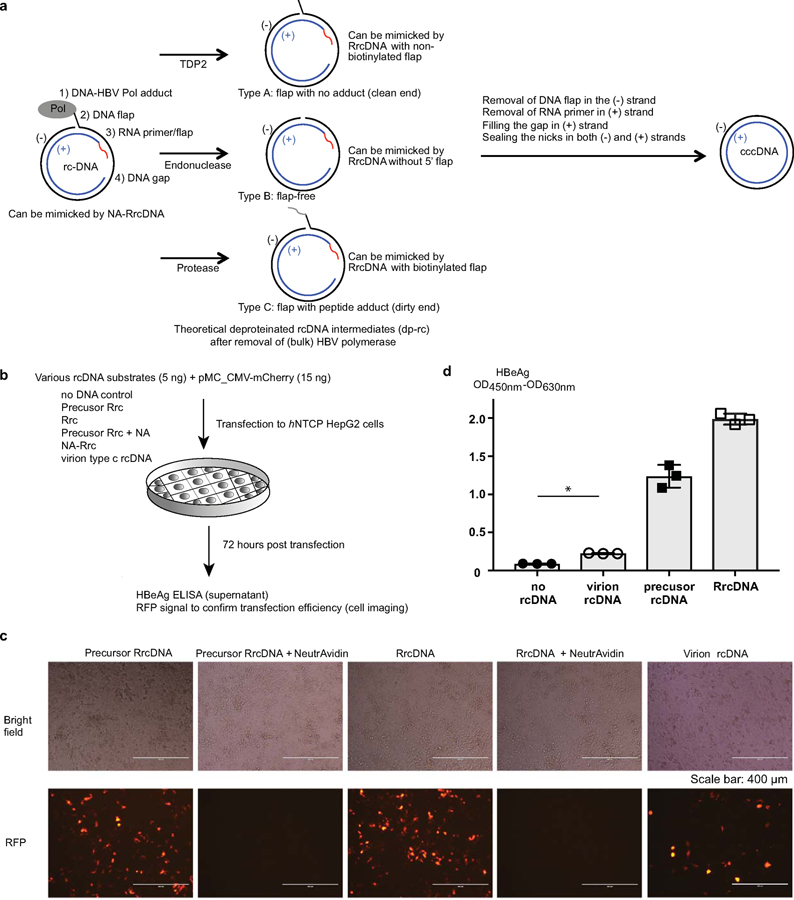
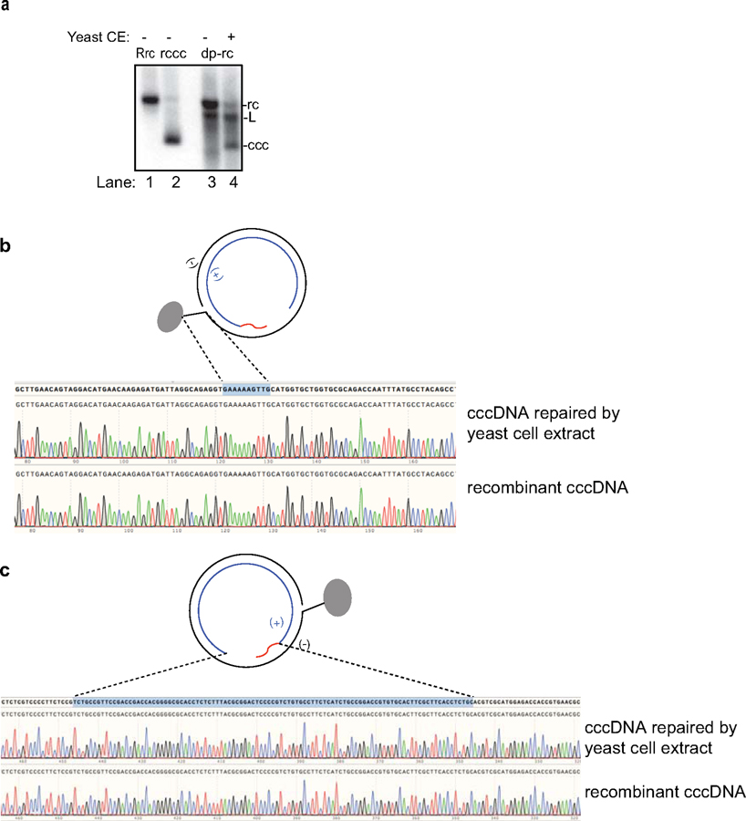
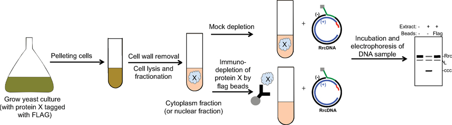
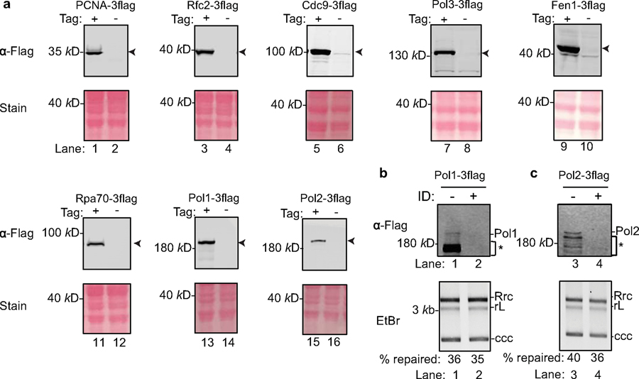
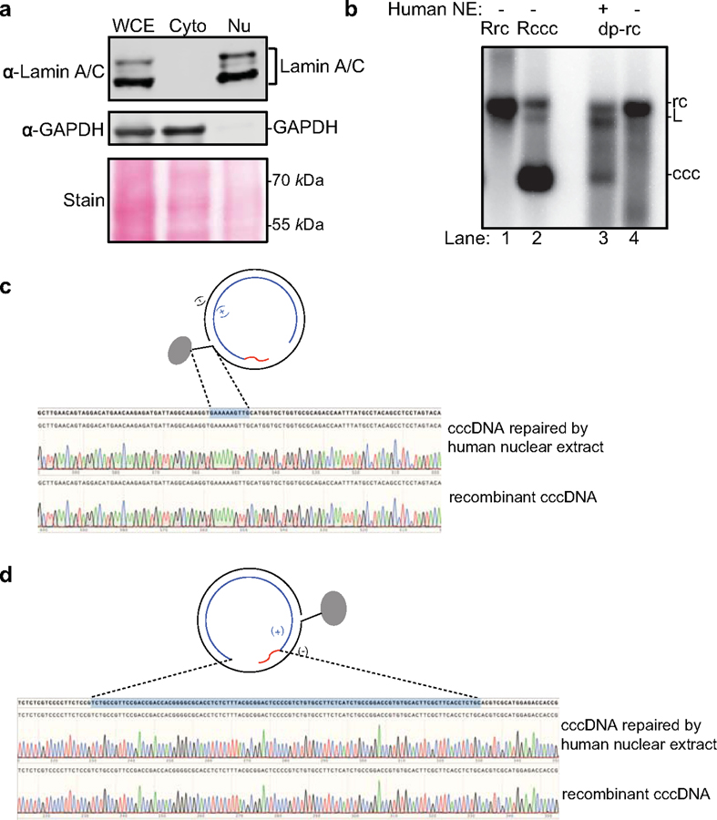
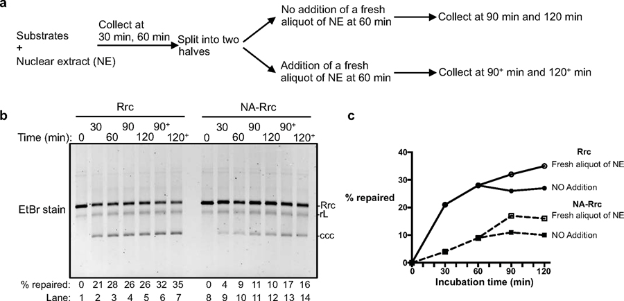
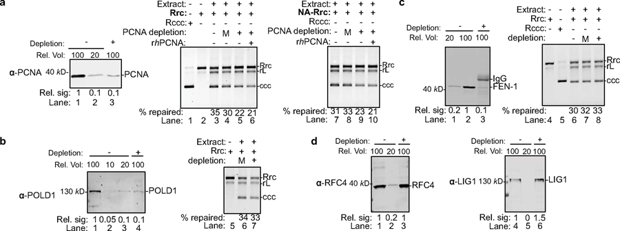
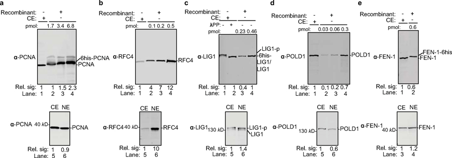
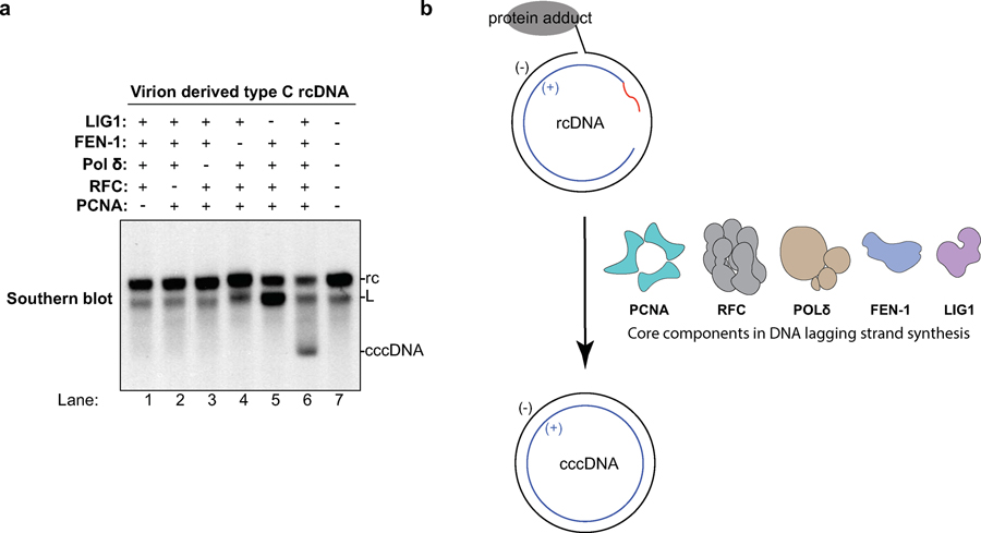
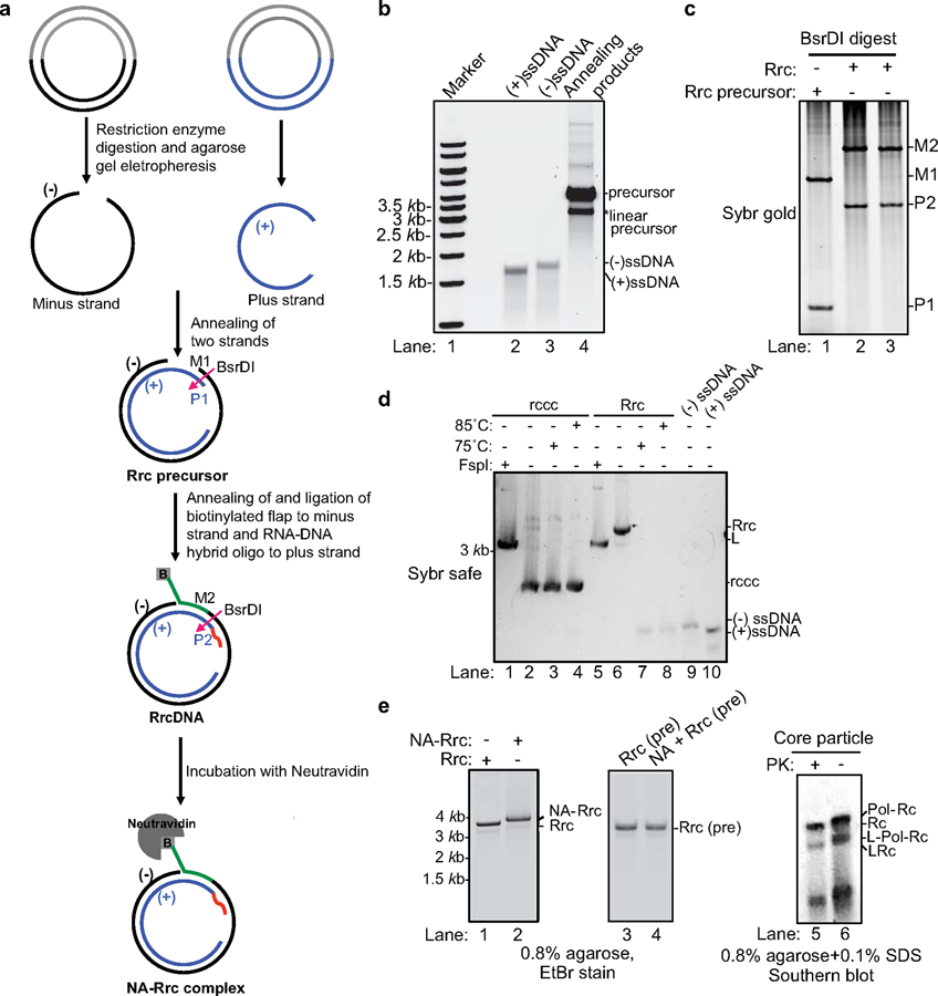
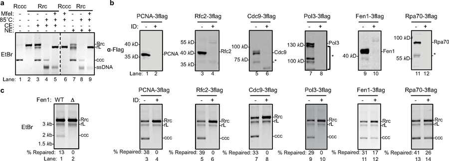
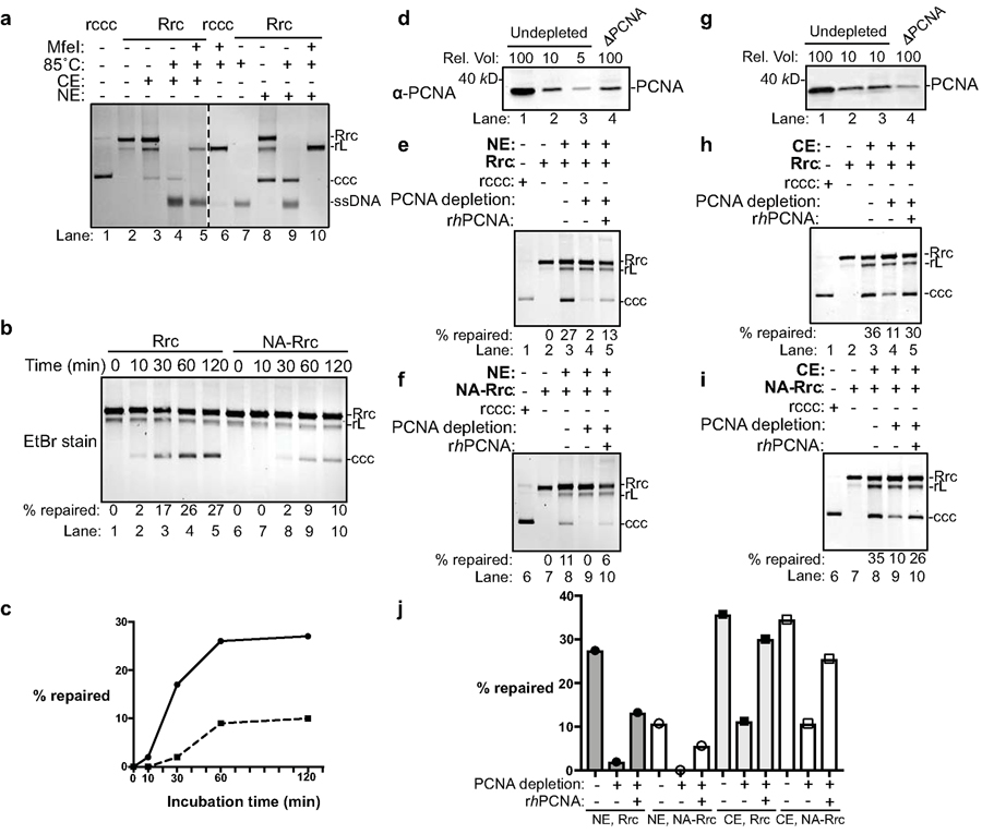
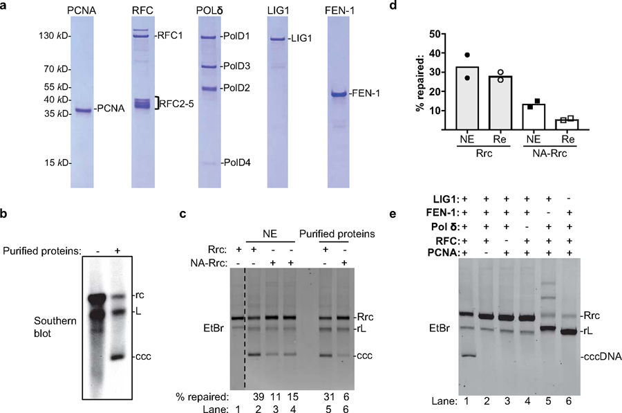
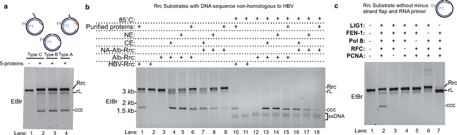
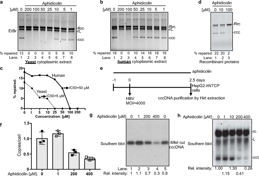
Chronic hepatitis B virus (HBV) infections result in 887,000 deaths annually. The central challenge in curing HBV is eradication of the stable covalently closed circular DNA (cccDNA) form of the viral genome, which is formed by the repair of lesion-bearing HBV relaxed circular DNA delivered by the virions to hepatocytes. The complete and minimal set of host factors involved in cccDNA formation is unknown, largely due to the lack of a biochemical system that fully reconstitutes cccDNA formation. Here, we have developed experimental systems where various HBV relaxed-circular-DNA substrates are repaired to form cccDNA by both cell extracts and purified human proteins. Using yeast- and human-extract screenings, we identified five core components of lagging-strand synthesis as essential for cccDNA formation: proliferating cell nuclear antigen, the replication factor C complex, DNA polymerase δ, flap endonuclease 1 and DNA ligase 1. We reconstituted cccDNA formation with purified human homologues, establishing these as a minimal set of factors for cccDNA formation. We further demonstrated that treatment with the DNA-polymerase inhibitor aphidicolin diminishes cccDNA formation both in biochemical assays and in HBV-infected human cells. Together, our findings define key components in HBV cccDNA formation.


Conflict of interest statement

Competing interests

The authors declare no competing interests.

Figures

Extended Data Fig. 1
Extended Data Fig. 1. Characteristics of HBV rcDNA and effects of transfection of various rcDNA substrates to human hepatoma cells.
(a) Four types of structures on HBV rcDNA need to be repaired by host repair factors to form cccDNA. Red line, a RNA primer/flap; Pol, HBV polymerase. A model describing the steps of rcDNA repair is as following: Removal of HBV Polymerase adduct from rcDNA can be achieved by TDP2, unknown endonuclease and protease digestion, generating 3 types (type A–type C) of deproteinated rcDNA (dp-rcDNA). Dp-rc DNA needs to be further processed to remove DNA flap and RNA primer/flap, fill the gap, and ligate the nicks. HBV rcDNA, and type A–type C dp-rcDNA can be mimicked by indicated recombinant rcDNA substrates (see Fig.1 for details) to study cccDNA formation in our biochemical repair system. (b–d) Examination of HBeAg production after transfection with X-tremeGENE™ reagent of various HBV rcDNA substrates (described in (a) and Fig. 1a) into hNTCP HepG2 cells. (b) Schematics of transfection experiments. 5 ng of individual control and rcDNA substrates together with 15 ng of mCherry expressing plasmids were transfected to hNTCP HepG2 cells seeded in a 96-well plate. 72 hours post transfection, transfection efficiency of each rcDNA substrates was estimated by mCherry fluorescence (c, scale bars indicate 400 μm) and HBeAg production in the supernatant was detected by ELISA (n=3, independent experiments) (d). Note that addition of NeutrAvidin strongly inhibited transfection efficiency shown in (c, compare 1st and 2nd panel). Statistic comparisons between ‘no rcDNA’ and ‘virion rcDNA’ samples were made using student-t test (two-tailed), *p value is 8×10⁻⁵. Bar value indicates mean of three measurements, and error bars are s.d. All data shown are representatives of three independent experiments.
Extended Data Fig. 2
Extended Data Fig. 2. Yeast cell extract fully supports repair of rcDNA to form cccDNA.
(a) Repair of HBV virion-derived deproteinated rcDNA to cccDNA in yeast cell extract. Repair products are analyzed by Southern blotting (see methods for details). Un-treated recombinant rcDNA (RrcDNA) (lane 1), recombinant cccDNA (RcccDNA) (lane 2), and virion-derived rcDNA (lane 3) were used as controls. Lane 4, repair product following incubation of rcDNA with yeast cell extract (CE). L, linearized rcDNA. (b–c) Both minus and plus strands in RrcDNA are faithfully repaired by yeast cytoplasmic extract to form cccDNA. (b) DNA sequences of the minus strand of repaired cccDNA (Fig. 2a, lane 3) and recombinant cccDNA. The 10 nt flap region on the minus strand of rcDNA is indicated by a blue shaded box and dashed lines. (c) DNA sequences of the plus strand of repaired cccDNA (Fig. 2a, lane 3) and recombinant cccDNA. The ssDNA gap and RNA primer-containing region on the plus strand of rcDNA is indicated by a blue shaded box and dashed lines. All data shown are representatives of two independent experiments.
Extended Data Fig. 3
Extended Data Fig. 3. Schematic of a screening system using yeast cell extract to identify host factors essential for HBV rc- to cccDNA conversion.
Culture of yeast strains with a given protein ‘X’ tagged with Flag epitope are grown and pelleted. Cell pellets are treated with zymolyase to remove the cell wall and subsequently lysed. The cell lysate is separated into either cytoplasmic or nuclear fractions. Flag-tagged protein X is either mock depleted or depleted with anti-Flag antibody-conjugated beads. Both cell extracts are then incubated with recombinant rcDNA (RrcDNA), and the DNA from the repair reactions is subsequently extracted and separated on an agarose gel and visualized by EtBr staining or Southern blotting. A hypothetical gel pattern is shown – repair of RrcDNA will lead to the appearance of a cccDNA band that is expected to migrate faster. L stands for linearized RrcDNA due to nicking and shearing.
Extended Data Fig. 4
Extended Data Fig. 4. Tagging of yeast replication and repair factors with a C-terminal Flag epitope and effects of immuno-depletion of Pol1 and Pol2 on cccDNA formation.
(a) Successful fusion of a 3x Flag tag at the endogenous chromosomal loci of various repair genes was confirmed by immunoblots using anti-Flag M2 antibody. Arrowheads indicate 3xFlag fusion gene products. Ponceau stains indicate equal loading. Repeated four times independently. (b) Depletion of Pol1 (top) does not affect cccDNA formation (bottom). (c) Immuno-depletion of Pol2 to undetectable levels (top) does not affect cccDNA formation (bottom). * indicates degraded proteins; ID stands for immunodepletion. Repeated twice independently.
Extended Data Fig. 5
Extended Data Fig. 5. Repair of rcDNA to form cccDNA using human nuclear extract.
(a) Total cell, cytoplasmic, and nuclear extract from human HepG2 hepatoma cells were analyzed by western blotting to confirm separation of cytoplasmic and nuclear contents. Nuclear factor Lamin A/C, and cytoplasmic factor GAPDH were detected by specific antibodies. Cyto, cytoplasmic fraction; Nu, nuclear fraction. (b) HBV virion-derived deproteinated rcDNA is repaired with human nuclear extract (NE) and cccDNA formation is analyzed by Southern blotting. Un-treated recombinant rcDNA (RrcDNA) (lane 1), recombinant cccDNA (RcccDNA) (lane 2), and virion-derived rcDNA (lane 4) were used as controls. Lane 3, repair product following incubation of rcDNA with human nuclear extract (NE). (c) DNA sequences of the minus strand of repaired cccDNA (extracted from Fig. 3a, lane 8) and recombinant cccDNA. The 10 nt flap region on the minus strand of rcDNA is indicated by a blue shaded box and dashed lines. (d) DNA sequences of the plus strand of repaired cccDNA (extracted from Fig. 3a, lane 8) and recombinant cccDNA. The ssDNA gap and RNA primer-containing region on the plus strand of rcDNA is indicated by a blue shaded box and dashed lines. All data shown are representatives of two independent experiments.
Extended Data Fig. 6
Extended Data Fig. 6. Replenishment of fresh nuclear extract at cccDNA formation plateau only marginally improves cccDNA formation.
Related to Fig. 3b–c. (a) Schematics showing a time course for cccDNA formation reaction in human nuclear extracts. At 60 min, the reaction solution was split into two parts; one part was supplemented with a fresh aliquot of nuclear extract, while the other was not. (b) Time course assay showing the kinetics of cccDNA formation from both RrcDNA (lanes 1–7) and NeutrAvidin (NA)-RrcDNA complex (lanes 8–14) as described in (a). Efficiency of cccDNA formation was calculated as in Fig. 2c and indicated in row ‘% repaired’. (c) The efficiency of cccDNA formation from (b) is plotted against incubation time. All data shown are representatives of two independent experiments.
Extended Data Fig. 7
Extended Data Fig. 7. 80% depletion of PCNA and POLD1 and 90% depletion of FEN-1 do not affect cccDNA formation in human cytoplasmic extracts.
(a) Depletion of 80% of PCNA in human cytoplasmic extract (left) does not affect repair of RrcDNA (middle) and NA-RrcDNA (21). Experiments were carried out as in Fig. 3g–i. M, Mock depletion using mouse IgG. (b–c) same as (a), except that POLD1 and FEN-1 are examined, respectively. M, Mock depletion using rabbit IgG. (d) Antibodies against the RFC4 subunit of the RFC complex and LIG1 fail to achieve protein depletion in human cytoplasmic extracts. (a–d) a relative volume (rel. vol) of 100 corresponds to 0.5 μl extract. All data shown are representatives of two independent experiments.
Extended Data Fig. 8
Extended Data Fig. 8. Estimation of the concentration of repair factors in human cell extract.
(a) The concentrations of PCNA in cytoplasmic extract (CE, top) and nuclear extract (NE, bottom) are compared by immunoblot with purified 6xhistidine-tagged PCNA of the indicated amount. 1 μl of extracts were contained in CE and NE. Relative signal of PCNA bands was calculated by setting that of cytoplasmic extract to 1. (b–e) Same as (a), except that RFC4, LIG1, POLD1, and FEN-1 are examined. LIG1-p, phosphorylated form of LIG1. λPP, lambda phosphatase. Note that λPP treatment (lane 2) in (c) reduces the mobility of phosphorylated LIG1 (lane 1) to that of the non-phosphorylated form (lanes 2–4). All data shown are representatives of two independent experiments.
Extended Data Fig. 9
Extended Data Fig. 9. All five factors are necessary and sufficient for repair of virion derived rcDNA and a summary of human factors involved in cccDNA formation in this study.
(a) The repair of virion derived type C rcDNA requires all five factors as recombinant rcDNA substrates used in Fig. 4e. (b) The core components involved in DNA lagging strand synthesis – PCNA, RFC, POLδ, FEN-1 and LIG1 – constitute a minimal set of factors for cccDNA formation in this study. All data shown are representatives of two independent experiments.
Extended Data Fig. 10
Extended Data Fig. 10. Strains and plasmids used in this study
Saccharomyces cerevisiae strains were isogenic to W1588–4C, a RAD5 derivative of W303 (MATa ade2–1 can1–100 ura3–1 his3–11,15 leu2–3,112 trp1–1 rad5–535) (27). Only one strain is listed for each genotype, but at least two independent isolates of each genotype were used in the experiments.
Fig. 1 |
Fig. 1 |. Generation of recombinant rcDNA (RrcDNA) and the NeutrAvidin RrcDNA (NA-RrcDNA) complex.
(a) Schematic representation of the generation of recombinant HBV rcDNA. RrcDNA and NA-RrcDNA are mimics of dp-rcDNA and HBV polymerase covalently linked rcDNA, respectively. BsrDI cleavage was used to monitor the efficiency of RrcDNA formation in (c) (BsrDI restriction sites are indicated by magenta arrows); green line, biotinylated flap; red line, RNA primer. Note that M1 and M2 are two oligos released from the minus strands of RrcDNA precursor and RrcDNA after BsrDI digestion, while P1 and P2 are two oligos released from the plus strands after BsrDI digestion. (b) Annealing products of recombinant minus and plus strand ssDNA to form RrcDNA precursor were monitored by SYBR™ safe staining. (c) Efficiency of RrcDNA generation is analyzed by formation of M2 and P2 oligos after BsrDI digestion and urea-PAGE gel electrophoresis followed by SYBR™ gold staining. Repeated three times independently. (d) Electrophoretic mobility of RrcDNA and recombinant cccDNA (rccc) after heat and restriction enzyme FspI treatment are compared on an agarose gel stained by SYBR™ safe. Repeated twice independently. (e) Both NA-RrcDNA (lanes 1–2) and the authentic HBV protein-rcDNA complexes (lanes 5–6) display a characteristic shift in mobility as compared to their deproteinated counterparts (5,17). NeutrAvidin does not cause non-specific mobility shift of precursor RrcDNA (lanes 3–4). PK, proteinase K treatment; L, linear; Pol, HBV polymerase. Lanes 1–4 were repeated independently three times; lanes 5–6 twice.
Fig. 2 |
Fig. 2 |. PCNA, RFC complex, Cdc9, Polδ, and Fen1 are required to repair recombinant rc-DNA to form cccDNA in yeast cell extracts.
(a) cccDNA formation assay (see methods for details) in yeast cytoplasmic (CE) and nuclear extracts (NE) were carried out to examine the repair of recombinant rcDNA (Rrc) to form cccDNA. Repair products were resolved on an agarose gel and stained with ethidium bromide (EtBr). Both yeast CE and NE fully support cccDNA formation (lanes 3 and 7). Untreated recombinant cccDNA (Rccc) and Rrc serve as controls (lanes 1–2). Repair products of CE (lanes 4–5) or NE (lanes 8–9) were treated with 85°C for 5 min alone or 85°C for 5 min and followed by MfeI digestion. Repaired cccDNA is heat resistant and shows mobility shift upon linearization. rL, recombinant linear DNA. Dashed line indicates removal of superfluous lanes. Repeated twice independently. (b) Near complete immuno-depletion of Flag-tagged-DNA repair proteins in yeast cell extracts examined by western blotting. ID, Immuno-depletion; *, indicates degradation bands. Repeated twice independently. (c) Depletion of factors involved in lagging strand synthesis abrogates repair of RrcDNA to form cccDNA. Lanes 1–2, cccDNA formation assay using extracts from WT or Fen1 null yeast strains. Lanes 3–14, same as lanes 1–2, except immunodepleted extracts from (b) are analyzed. % repaired, the percentage of total RrcDNA that is repaired to form cccDNA was calculated by the intensity of the ccc band divided by the sum intensities of Rrc, rL and ccc bands. Absolute values are shown above each lane number. Repeated twice independently.
Fig. 3 |
Fig. 3 |. PCNA is required for cccDNA formation in human cell extracts.
(a) Cytoplasmic and nuclear extracts from hNTCP-expressing HepG2 cells fully support cccDNA formation. Repair of RrcDNA was carried out and analyzed as in Fig. 2a, except the incubation temperature was 37°C instead of 30°C. Dashed line indicates removal of superfluous lanes. (b) Time course assay showing the kinetics of cccDNA formation from both RrcDNA (lanes 1–5) and NA-RrcDNA complex (lanes 6–10) in nuclear extracts. Experiments were performed as in (a), except that reactions were terminated at indicated time points. Efficiency of cccDNA formation was calculated as in Fig. 2c and indicated in row ‘% repaired’. (c) The efficiency of cccDNA formation from (b) is plotted against incubation time. (d) Immuno-depletion of more than 90% of PCNA from human nuclear extracts. Un-depleted and PCNA-depleted extracts were analyzed by western blotting using an anti-PCNA antibody. A relative volume (rel. vol) of 100 corresponds to 0.5 μl extract. Depletion of PCNA drastically reduced repair efficiency of RrcDNA (e) and NA-RrcDNA (f) in human nuclear extracts. Untreated recombinant cccDNA (Rccc) and Rrc were used as control (lanes 1, 2). Lanes 3 and 8, mock-depleted extract with mouse IgG; lanes 4 and 9, PCNA-depleted extract from (d) was used; lanes 5 and 10, addition of recombinant human PCNA (rhPCNA, shown in Fig. 4a) to PCNA-depleted extract restored cccDNA repair. (g–i) same to (d–f), except that human cytoplasmic extracts were used. (j) The efficiency of cccDNA formation from (e–f, g–i) is plotted. All data shown were repeated twice independently.
Fig. 4 |
Fig. 4 |. Purified human PCNA, RFC, LIG1, POLδ, and FEN-1 define the minimal set of factors for cccDNA formation.
(a) Purified human proteins involved in DNA lagging strand synthesis analyzed by SDS-PAGE with Coomassie staining. Repeated three times independently. (b) cccDNA formation assay using virion derived dp-rcDNA and purified human proteins from (a). Repeated twice independently. (c) The repair efficiency of RrcDNA and NA-RrcDNA by human nuclear extract (lanes 2–4) versus by purified human proteins (lanes 5–6) were compared. Untreated RrcDNA was used as a control (lane 1). Dashed line indicates removal of superfluous lanes. Repeated three times independently. (d) The repair efficiency from (c) is plotted (n=2). NE, nuclear extract. PP, purified proteins. Bar value indicates mean of two measurements. (e) All five factors are required for cccDNA formation. Omission of factors is indicated by ‘–’. All data presented are representatives of two to three independent experiments. Repeated twice independently.
Fig. 5 |
Fig. 5 |. Repair of RrcDNA with various lesion structure and sequences with purified proteins and human cell extracts.
(a) Analogous to RrcDNA that mimics type C dp-rcDNA (lane 2), RrcDNA substrates that mimic type A (no flap) and type B dp-rcDNA (clean-ended flap) can also be repaired by 5 purified human factors (lanes 3–4). Repeated twice independently. (b) A 3.2 kb RrcDNA or NA-RrcDNA containing a flap and RNA primer, and with all HBV sequence replaced by albumin promoter and mCherry sequences, was subject to cccDNA formation reaction in human cytoplasmic extract (lanes 4, 7), nuclear extract (lanes 5, 8), and purified protein system (lanes 6, 9). HBV-RrcDNA was used as a control (lane 1). The cccDNA reaction products from lanes 1–9 were subjected to heat treatment at 85°C for 5 min and loaded to lanes 10–18 to show cccDNA bands. Alb: human Albumin and mCherry sequences; NA: NeutrAvidin. Repeated twice independently. (c) Rrc Substrate without DNA flap and RNA primer/flap was incubated with 5 purified human factors (lane 2) or with factors omitted (lanes 3–7), and cccDNA formation was detected by agarose gel electrophoresis. All data presented are representatives of two to three independent experiments. Repeated twice independently.
Fig. 6 |
Fig. 6 |. DNA polymerase inhibitor aphidicolin inhibits cccDNA formation in cell extracts, in the purified protein system, and HBV-infected human hepatoma cells.
Robust inhibition of cccDNA formation by aphidicolin in yeast ((a), at 30°C, protein concentration ~15 mg/ml) and human HepG2-hNTCP cytoplasmic extract ((b), at 37°C, protein concentration ~30 mg/ml). (c) Efficiencies of cccDNA formation from (a) and (b) are plotted against aphidicolin concentration. (d) Aphidicolin inhibits cccDNA formation by the purified protein components (at 37°C). (e) Schematic for testing the effect of aphidicolin on cccDNA formation in hNTCP-expressing HepG2 cells infected with HBV. ‘–1’ indicates aphidicolin treatment started one day before HBV challenge. MOI, multiplicity of infection. (f) ExoI/ExoIII/T5 nuclease treated cccDNA copy numbers from HBV-infected cells detected by quantitative PCR (n=3). Bar value indicates mean of three measurements, and error bars are s.d. (g) cccDNA samples from (f) were linearized by MfeI and analyzed by Southern blotting. The relative intensities of cccDNA bands were normalized to those of mock-treated cells (lane 1). (h) Southern blotting of cccDNA extracted by Hirt method without ExoI/ExoIII/T5 nuclease treatment. Southern blotting was performed as in (g). All data presented are representatives of two to three independent experiments. All data shown [except for (f, n=3)] are repeated twice independently.

Comment in

References

    1. Winer BY & Ploss A. Determinants of hepatitis B and delta virus host tropism. Curr Opin Virol 13, 109–116 (2015). - PMC - PubMed
    1. Nassal M. HBV cccDNA: viral persistence reservoir and key obstacle for a cure of chronic hepatitis B. Gut 64, 1972–1984 (2015). - PubMed
    1. Yan H. et al. Sodium taurocholate cotransporting polypeptide is a functional receptor for human hepatitis B and D virus. Elife 1, e00049 (2012). - PMC - PubMed
    1. Wang GH & Seeger C. The reverse transcriptase of hepatitis B virus acts as a protein primer for viral DNA synthesis. Cell 71, 663–670 (1992). - PubMed
    1. Gerlich WH & Robinson WS. Hepatitis B virus contains protein attached to the 5’ terminus of its complete DNA strand. Cell 21, 801–809 (1980). - PubMed

Publication types