

Pulmonary manifestations of systemic karyomegaly
- PMID: 32154101
- PMCID: PMC7058920
- DOI: 10.1016/j.rmcr.2020.101032
Pulmonary manifestations of systemic karyomegaly
Abstract
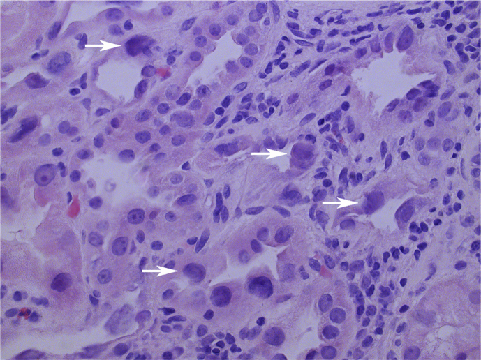
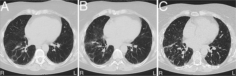
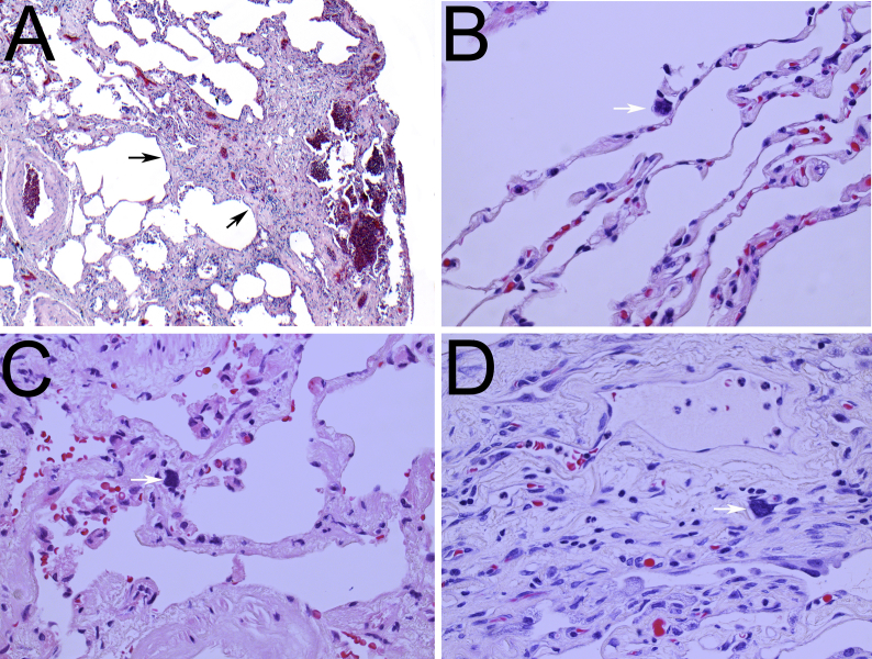
Over 40 years ago, abnormal enlargement of the nucleus of tubular epithelial cells was reported in a rare distinct hereditary chronic interstitial nephritis, karyomegalic interstitial nephritis (KIN). Here, we report the second case of systemic karyomegaly with pulmonary manifestations and present a detailed characterization of the karyomegalic cells in lung parenchyma. A 59-year-old woman who was diagnosed with KIN developed renal failure and eventually received a renal transplant later evaluated for chronic and progressive restrictive lung disease. The KIN diagnosis prompted us to carefully examine her lung parenchyma. Karyomegalic cells were identified in the alveolar epithelium, interstitium, as well as, in the vascular wall. Viral serological and biochemical blood analyses were negative. We consider that the pulmonary manifestations of karyomegaly expands the differential diagnosis of interstitial lung disease in patients with KIN.
Keywords: Interstitial lung disease; Karyomegalic interstitial nephritis; Karyomegaly.
© 2020 The Authors.
Conflict of interest statement
None declared.
Figures
References
-
- Burry A.F. Extreme dysplasia in renal epithelium of a young woman dying from hepatocarcinoma. J. Pathol. 1974;113(3):147–150. - PubMed
-
- Sclare G. A case of unexplained karyomegaly. Beitr. Pathol. 1976;157(3):301–306. - PubMed
-
- Mihatsch M.J. Systemic karyomegaly associated with chronic interstitial nephritis. A new disease entity? Clin. Nephrol. 1979;12(2):54–62. - PubMed
