. 2020 Feb 21:8:110.
doi: 10.3389/fbioe.2020.00110. eCollection 2020.

Auto Micro Atomization Delivery of Human Epidermal Organoids Improves Therapeutic Effects for Skin Wound Healing

Affiliations

Auto Micro Atomization Delivery of Human Epidermal Organoids Improves Therapeutic Effects for Skin Wound Healing

Mingyang Chang et al. Front Bioeng Biotechnol. .

Abstract

Severe skin wounds are often associated with large areas of damaged tissue, resulting in substantial loss of fluids containing electrolytes and proteins. The net result is a vulnerability clinically to skin infections. Therapies aiming to close these large openings are effective in reducing the complications of severe skin wounds. Recently, cell transplantation therapy showed the potential for rapid re-epithelialization of severe skin wounds. Here, we show the improved effects of cell transplantation therapy using a robust protocol of efficient expansion and delivery of epidermal cells for treatment of severe skin wounds. Human skin tissues were used to generate human epidermal organoids maintained under newly established culture conditions. The human epidermal organoids showed an improved capacity of passaging for at least 10 rounds, enabling organoids to expand to cell numbers required for clinical applications. A newly designed auto micro-atomization device (AMAD) was developed for delivery of human epidermal organoids onto the sites of severe skin wounds enhancing uniform and concentrated delivery of organoids, facilitating their engraftment and differentiation for skin reconstitution. With the optimal design and using pneumatic AMAD, both survival and functions of organoids were effectively protected during the spraying process. Cells in the sprayed human epidermal organoids participated in the regeneration of the epidermis at wound sites in a mouse model and accelerated wound healing significantly. The novel AMAD and out new protocol with enhanced effects with respect to both organoid expansion and efficient transplantation will be used for clincal treatments of complex, uneven, or large-area severe skin wounds.

Keywords: auto micro atomization device (AMAD); cell delivery; cell therapy; organoids; wound healing.


Figures

Figure 1
Figure 1
The potenial therapeutical application of the cell-delivery system for treatment of large and irregular wounds in clinic by an AMAD. In this study, primary human skin epidermal organoids were sprayed and delivered onto the wound. They effectively accelerated skin wound healing through participation in epidermis regeneration.
Figure 2
Figure 2
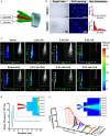
Fluid dynamics analysis of the AMAD. (A) The structural and working principle diagram of the nozzle, including the liquid and gas outlet. (B) Bright field and fluorescent images showing the droplets sprayed by the AMAD and injector and their size distribution. (C) The velocity distribution of different liquid at a series of viscosity, which analyzed from the laser-triggered high-speed photography (inside the frame). (D) The gas static pressure distribution of nozzle and outlet jet (inside the frame), and the distribution of gas static pressure on the central axis. (E) The gas velocity distribution of nozzle and outlet jet (inside the frame), and the axial and radial distribution of gas velocity after spraying.
Figure 3
Figure 3
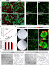
Cell survival and proliferation following spraying with the AMAD. (A) Live/Dead staining of HaCaT cells cultured for 2 and 12 h after seeding via pipette (negative control) vs. via spray. (B) The proliferation curve of HaCaT cells after seeding via pipette vs. spray. (C) The representative Live/Dead staining of HaCaT cells suspended and sprayed in different media containing Matrigel. (D) The bright field and fluorescent images of HaCaT organoids in Matrigel after being delivered by spray vs. pipette; the numbers in the photos indicate the percentages of live cells in the organoids. (E) The tubular network formation by HUVEC cells after being delivered by spray vs. pipette is noted along with information on the extracted skeletons of tubular networks analyzed by ImageJ (F) and with the parameters, including identified segments, nodes and meshes, of tube formation.
Figure 4
Figure 4
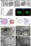
Characterization of an in vitro expanded epithelial cell organoid. (A) Representative serial images of an organoid growing at the indicated time points. Magnifications: 20 × [days 0, 3, 5, 7, passage (P) 1, P4 and P6]. Scale bar, 50 μm. (B) Live/Dead staining of skin organoids at P1 and P6. (C) The growth curve of skin organoids generated from isolated adult foreskin cells. (D) TEM analysis for skin organoids. The yellow arrows are used to indicate gap junctions or desmosomes (E) and tight junctions (TJ).
Figure 5
Figure 5
Human skin-derived organoids delivered by the AMAD improved wound healing. (A) Representative images of the mouse wound splinting model after spraying of the skin organoids; the medium without cells was used as the control group. (B) Analysis of wound size (%) of each group, and the wound area over time were measured as a percent of the original area, n = 3. (C) Wound sections on 7, 14, and 21 days after spraying were stained with H&E for general observation of skin layers. (D) Immunohistochemical staining of Ki67, CK14, and CK10 on the skin wound treated by spraying of epidermal organoids vs. control (saline).
Figure 6
Figure 6
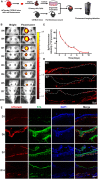
In vivo imaging to monitor the sprayed td-Tomato mice-derived organoids on full thickness wounds. (A) Schematic to demonstrate the normal mice with wounds, used to monitor the effects of tdTomato-autologous cells isolated from syngeneic mice. (B) Overlay of the representative light and fluorescent images of skin wounds monitored from day 0 to 10 after spraying organoids (“sprayed” on the left) on the wound bed. The wounds without sprayed organoids (“non-sprayed” on the right) were used to normalized the fluorescence. (C) The estimates of the presence of the donor cells in the wound bed were calculated by subtracting the level of “non-sprayed” from that of “sprayed” areas, n = 3. (D) Fluorescent imaging on frozen sections to detect the existence of tdTomato-autologous cells after spraying of donor cells on wound sites. (E) Immunofluorescence staining of the wound tissue at days 3, 5, 7, and 14 to indicate the integration of sprayed donor cells onto the wound beds. The tissues were stained for CK14.

Similar articles

Cited by

References

    1. Auxenfans C., Menet V., Catherine Z., Shipkov H., Lacroix P., Bertin-Maghit M., et al. . (2015). Cultured autologous keratinocytes in the treatment of large and deep burns: a retrospective study over 15 years. Burns 41, 71–79. 10.1016/j.burns.2014.05.019 - DOI - PubMed
    1. Bahoric A., Harrop A. R., Clarke H. M., Zuker R. M. (1997). Aerosol vehicle for delivery of epidermal cells – an in vitro study. Can. J. Plast. Surg. 5, 153–156. 10.1177/229255039700500301 - DOI
    1. Beneke V., Kuster F., Neehus A. L., Hesse C., Lopez-Rodriguez E., Haake K., et al. . (2018). An immune cell spray (ICS) formulation allows for the delivery of functional monocyte/macrophages. Sci. Rep. 8:16281. 10.1038/s41598-018-34524-2 - DOI - PMC - PubMed
    1. Chen P., Parks W. C. (2009). Role of matrix metalloproteinases in epithelial migration. J. Cell. Biochem. 108, 1233–1243. 10.1002/jcb.22363 - DOI - PMC - PubMed
    1. Cohen M., Bahoric A., Clarke H. M. (2001). Aerosolization of epidermal cells with fibrin glue for the epithelialization of porcine wounds with unfavorable topography. Plast. Reconstr. Surg. 107, 1208–1215. 10.1097/00006534-200104150-00018 - DOI - PubMed

LinkOut - more resources