Review
. 2020 Jul 1;116(8):1410-1423.
doi: 10.1093/cvr/cvaa034.

RUNX1: an emerging therapeutic target for cardiovascular disease

Affiliations
Review

RUNX1: an emerging therapeutic target for cardiovascular disease

Alexandra Riddell et al. Cardiovasc Res. .

Abstract

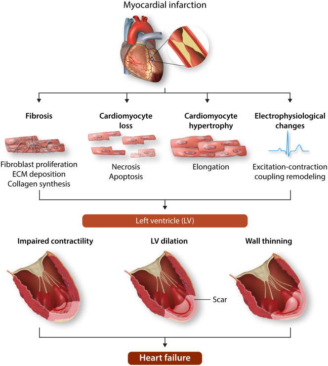
Runt-related transcription factor-1 (RUNX1), also known as acute myeloid leukaemia 1 protein (AML1), is a member of the core-binding factor family of transcription factors which modulate cell proliferation, differentiation, and survival in multiple systems. It is a master-regulator transcription factor, which has been implicated in diverse signalling pathways and cellular mechanisms during normal development and disease. RUNX1 is best characterized for its indispensable role for definitive haematopoiesis and its involvement in haematological malignancies. However, more recently RUNX1 has been identified as a key regulator of adverse cardiac remodelling following myocardial infarction. This review discusses the role RUNX1 plays in the heart and highlights its therapeutic potential as a target to limit the progression of adverse cardiac remodelling and heart failure.

Keywords: Adverse cardiac remodelling; Calcium; Cardiovascular diseases; Excitation–contraction coupling; Heart failure; Myocardial infarction; Runx1.


Figures

Figure 1
Figure 1
Pathological ventricular remodelling. Schematic showing remodelling of the left ventricle (LV) following myocardial infarction (MI). Following MI, a fibrosous scar forms in the infarcted tissue accompanied by myocyte loss. Adjacent to the infarcted tissue in the region bordering the remote LV, the border-zone region, myocytes thicken and elongate and excitation–contraction coupling becomes impaired. In terms of gross architecture of the LV, the early phase of remodelling is characterized by thinning and elongation of the infarcted zone followed by ventricular dilation, where the LV transitions from an elliptical shape to a spherical shape. Schematic was prepared using Servier Medical Art by Servier under a Creative Commons Attribution 3.0 Unported License.
Figure 2
Figure 2
RUNX1 structure and regulation. Illustration of RUNX1 transcriptional regulatory complexes for activation and repression of gene expression (A). Schematic depicting RUNX1 gene structure (B). Expression of RUNX1 is initiated by two promoters: distal P1 and proximal P2. (C) Alternative promoters and gene-splicing results in different 5ʹ-UTRs. The P1-5ʹ-UTR contains four exons and the P2-5ʹ-UTR has a single exon and an internal ribosome entry site (IRES). This results in three major isoforms of RUNX1; 1A and 1B transcribed from P2 and isoform 1C transcribed from P1. (D) Schematic of the proteins encoded by the RUNX1 isoforms with the major functional domains marked: runt-homology domain (RHD) and transactivation domain (TAD). Figure prepared using information from various sources.
Figure 3
Figure 3
Post-translational modifications of human RUNX1. Schematic depicting post-translational modifications of RUNX1b. Runt-homology domain (RHD) is the DNA- and CBFβ-binding domain. Transactivation domain (TAD) is important for transcriptional activation. Numbers refer to amino acid residues from N terminus. Ac, acetylation; APC, anaphase-promoting complex; K, lysine; Me, methylation; P, phosphorylation; R, arginine; S, serine; SCF, Skp1/Cullin/F-box protein complex; T, threonine; Ub, ubiquitination; Y, tyrosine. Figure prepared using information from various sources.,

References

    1. Conrad N, Judge A, Tran J, Mohseni H, Hedgecott D, Crespillo AP, Allison M, Hemingway H, Cleland JG, McMurray JJV, Rahimi K.. Temporal trends and patterns in heart failure incidence: a population-based study of 4 million individuals. Lancet 2018;391:572–580. - PMC - PubMed
    1. Dewan P, Rørth R, Jhund PS, Ferreira JP, Zannad F, Shen L, Køber L, Abraham WT, Desai AS, Dickstein K, Packer M, Rouleau JL, Solomon SD, Swedberg K, Zile MR, McMurray JJV.. Income inequality and outcomes in heart failure: a global between-country analysis. JACC Heart Fail 2019;7:336–346. - PubMed
    1. Konstam MA, Kramer DG, Patel AR, Maron MS, Udelson JE.. Left ventricular remodeling in heart failure: current concepts in clinical significance and assessment. JACC Cardiovasc Imaging 2011;4:98–108. - PubMed
    1. Burchfield JS, Xie M, Hill JA.. Pathological ventricular remodeling: mechanisms: part 1 of 2. Circulation 2013;128:388–400. - PMC - PubMed
    1. Cokkinos DV, Belogianneas C.. Left ventricular remodelling: a problem in search of solutions. Eur Cardiol 2016;11:29–35. - PMC - PubMed

Publication types

MeSH terms

Substances