

Effects of the probiotic formulation SLAB51 in in vitro and in vivo Parkinson's disease models
- PMID: 32155131
- PMCID: PMC7093198
- DOI: 10.18632/aging.102927
Effects of the probiotic formulation SLAB51 in in vitro and in vivo Parkinson's disease models
Abstract
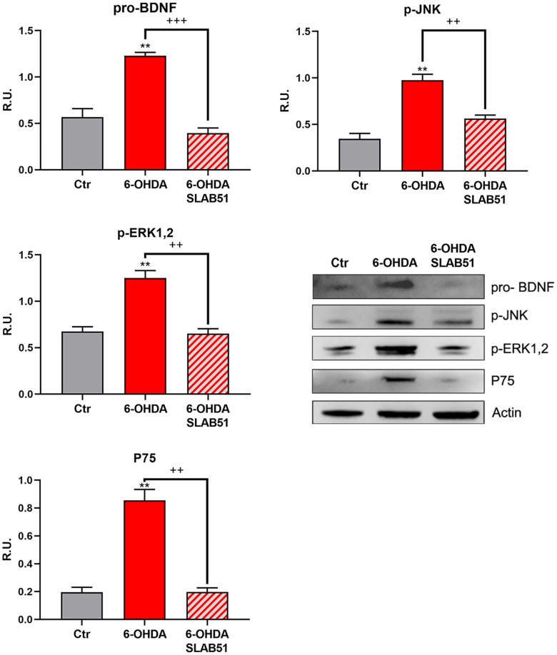
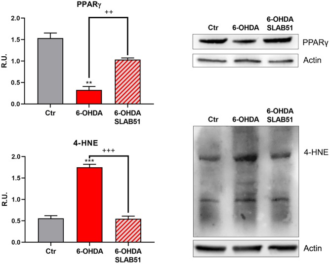
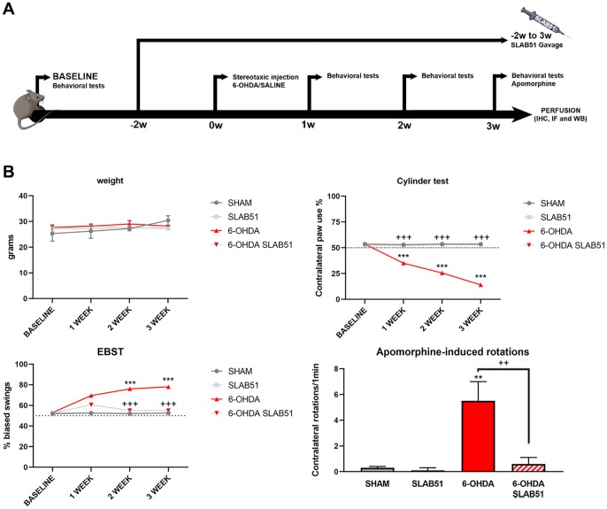
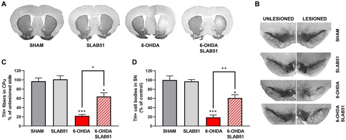
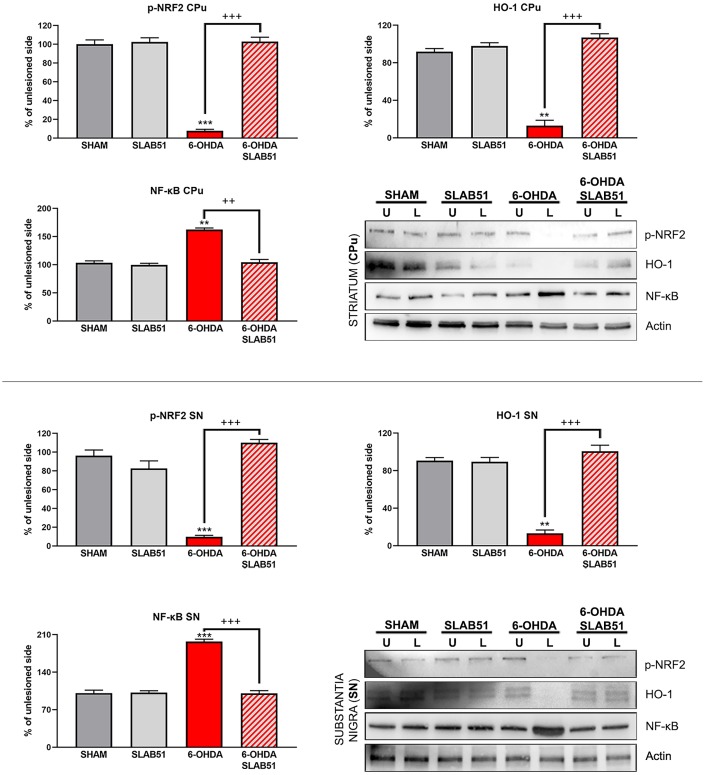
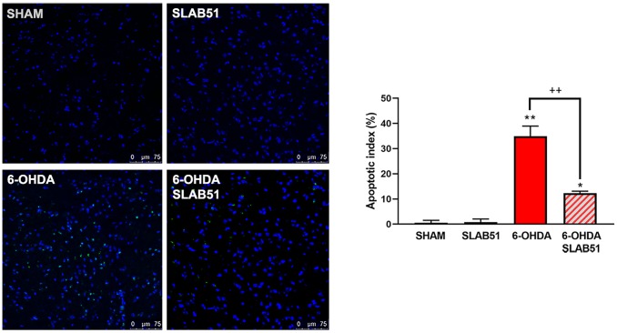
Parkinson is a common neurodegenerative disorder, characterized by motor and non-motor symptoms, including abnormalities in the gut function, which may appear before the motor sign. To date, there are treatments that can help relieve Parkinson' disease (PD)-associated symptoms, but there is no cure to control the onset and progression of this disorder. Altered components of the gut could represent a key role in gut-brain axis, which is a bidirectional system between the central nervous system and the enteric nervous system. Diet can alter the microbiota composition, affecting gut-brain axis function. Gut microbiome restoration through selected probiotics' administration has been reported. In this study, we investigated the effects of the novel formulation SLAB51 in PD. Our findings indicate that this probiotic formulation can counteract the detrimental effect of 6-OHDA in vitro and in vivo models of PD. The results suggest that SLAB51 can be a promising candidate for the prevention or as coadjuvant treatment of PD.
Keywords: 6-OHDA; BDNF; Parkinson's disease; probiotics; tyrosine hydroxylase.
Conflict of interest statement
Figures
Similar articles
-
Are We What We Eat? Impact of Diet on the Gut-Brain Axis in Parkinson's Disease.Nutrients. 2022 Jan 17;14(2):380. doi: 10.3390/nu14020380. Nutrients. 2022. PMID: 35057561 Free PMC article. Review.
-
Microbiome-Gut-Brain Axis and Toll-Like Receptors in Parkinson's Disease.Int J Mol Sci. 2018 Jun 6;19(6):1689. doi: 10.3390/ijms19061689. Int J Mol Sci. 2018. PMID: 29882798 Free PMC article. Review.
-
Research Progress of Microbiota-Gut-Brain Axis in Parkinson's Disease.J Integr Neurosci. 2023 Oct 30;22(6):157. doi: 10.31083/j.jin2206157. J Integr Neurosci. 2023. PMID: 38176929 Review.
-
Mind-altering with the gut: Modulation of the gut-brain axis with probiotics.J Microbiol. 2018 Mar;56(3):172-182. doi: 10.1007/s12275-018-8032-4. Epub 2018 Feb 28. J Microbiol. 2018. PMID: 29492874 Review.
-
The emerging role of probiotics in neurodegenerative diseases: new hope for Parkinson's disease?Neural Regen Res. 2021 Apr;16(4):628-634. doi: 10.4103/1673-5374.295270. Neural Regen Res. 2021. PMID: 33063712 Free PMC article.
Cited by
-
Agathobaculum butyriciproducens Shows Neuroprotective Effects in a 6-OHDA-Induced Mouse Model of Parkinson's Disease.J Microbiol Biotechnol. 2022 Sep 28;32(9):1168-1177. doi: 10.4014/jmb.2205.05032. Epub 2022 Sep 5. J Microbiol Biotechnol. 2022. PMID: 36168204 Free PMC article.
-
Neurotrophic factor-based pharmacological approaches in neurological disorders.Neural Regen Res. 2023 Jun;18(6):1220-1228. doi: 10.4103/1673-5374.358619. Neural Regen Res. 2023. PMID: 36453397 Free PMC article. Review.
-
The gut microbiota-brain axis in behaviour and brain disorders.Nat Rev Microbiol. 2021 Apr;19(4):241-255. doi: 10.1038/s41579-020-00460-0. Epub 2020 Oct 22. Nat Rev Microbiol. 2021. PMID: 33093662 Review.
-
When the microbiome helps the brain-current evidence.CNS Neurosci Ther. 2023 Jun;29 Suppl 1(Suppl 1):43-58. doi: 10.1111/cns.14076. Epub 2023 Jan 4. CNS Neurosci Ther. 2023. PMID: 36601680 Free PMC article. Review.
-
Neurodegenerative and Neurodevelopmental Diseases and the Gut-Brain Axis: The Potential of Therapeutic Targeting of the Microbiome.Int J Mol Sci. 2023 May 31;24(11):9577. doi: 10.3390/ijms24119577. Int J Mol Sci. 2023. PMID: 37298527 Free PMC article. Review.
References
-
- Castelli V, Benedetti E, Antonosante A, Catanesi M, Pitari G, Ippoliti R, Cimini A, d’Angelo M. Neuronal Cells Rearrangement During Aging and Neurodegenerative Disease: Metabolism, Oxidative Stress and Organelles Dynamic. Front Mol Neurosci. 2019; 12:132. 10.3389/fnmol.2019.00132 - DOI - PMC - PubMed
-
- Rojas D, Della Pelle F, Del Carlo M, d’Angelo M, Dominguez-Benot R, Cimini A, Escarpa A, Compagnone D. Electrodeposited Prussian Blue on carbon black modified disposable electrodes for direct enzyme-free H2O2 sensing in a Parkinson’s disease in vitro model. Sens Actuators B Chem. 2018; 275:402–08. 10.1016/j.snb.2018.08.040 - DOI
MeSH terms
LinkOut - more resources
Full Text Sources
