. 2020 Mar 10;15(3):e0230001.
doi: 10.1371/journal.pone.0230001. eCollection 2020.

Practical adoption of state-of-the-art hiPSC-cardiomyocyte differentiation techniques

Affiliations

Practical adoption of state-of-the-art hiPSC-cardiomyocyte differentiation techniques

Cassady E Rupert et al. PLoS One. .

Abstract

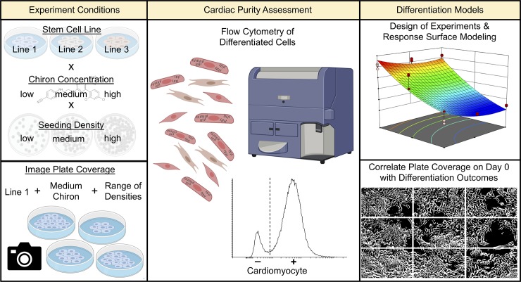
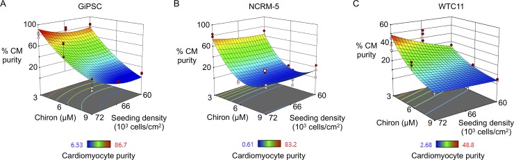
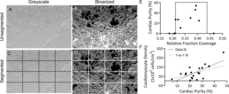
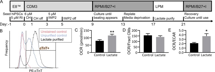
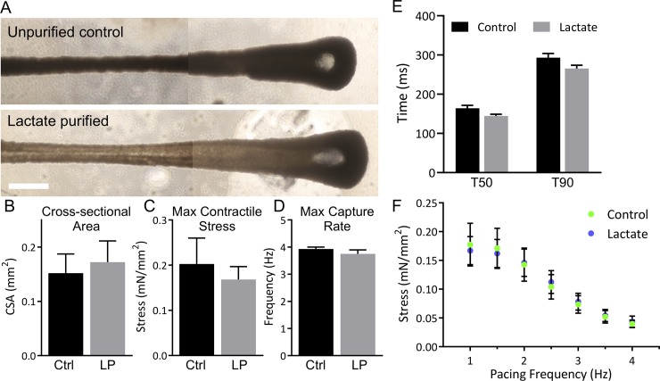
Human induced pluripotent stem cell (hiPSC)-derived cardiomyocytes are a valuable resource for cardiac therapeutic development; however, generation of these cells in large numbers and high purity is a limitation in widespread adoption. Here, design of experiments (DOE) is used to investigate the cardiac differentiation space of three hiPSC lines when varying CHIR99027 concentration and cell seeding density, and a novel image analysis is developed to evaluate plate coverage when initiating differentiation. Metabolic selection via lactate purifies hiPSC-cardiomyocyte populations, and the bioenergetic phenotype and engineered tissue mechanics of purified and unpurified hiPSC-cardiomyocytes are compared. Findings demonstrate that when initiating differentiation one day after hiPSC plating, low (3 μM) Chiron and 72 x 103 cells/cm2 seeding density result in peak cardiac purity (50-90%) for all three hiPSC lines. Our results confirm that metabolic selection with lactate shifts hiPSC-cardiomyocyte metabolism towards oxidative phosphorylation, but this more "mature" metabolic phenotype does not by itself result in a more mature contractile phenotype in engineered cardiac tissues at one week of culture in 3D tissues. This study provides widely adaptable methods including novel image analysis code and parameters for refining hiPSC-cardiomyocyte differentiation and describes the practical implications of metabolic selection of cardiomyocytes for downstream tissue engineering applications.


Conflict of interest statement

The authors have declared that no competing interests exist.

Figures

Fig 1
Fig 1. Schematic of differentiation optimization experiments.
Left column: Design of experiments (DOE) studies were performed using three hiPSC lines (GiPSC, NCRM-5, WTC11), three Chiron concentrations (3, 6, 9 μM), and three seeding densities (60, 66, 72 x103 cells/cm2; top). Image analysis studies were performed by imaging one hiPSC line (GiPSC) with one Chiron concentration (6 μM) at the beginning of differentiation across a range of seeding densities (bottom). Middle column: Output cell populations of all studies were analyzed by flow cytometry to identify percentage of cardiomyocytes by expression of cardiac troponin T. Right column: Cardiomyocyte purity was modeled as a function of seeding density and Chiron concentration (top). Phase contrast images of cells at the beginning of differentiation were correlated with cardiomyocyte purity through automated image analysis in Matlab (bottom).
Fig 2
Fig 2. Chiron concentration and seeding density uniquely affect cardiac differentiation outcome.
DOE (design of experiments) surface plots of directed differentiation outcomes in human induced pluripotent stem cell lines obtained from commercial (GiPSC; A), national bank (NCRM-5; B), and research institution (WTC11; C) sources. Red and pink circles indicate design points above and below predicted values, respectively. N = 9 per group per condition from 3 biological replicates (i.e., batches of hiPSC-cardiomyocytes).
Fig 3
Fig 3. Segmented thresholding shows range of plate coverage for cardiomyocyte differentiation using 6 μM Chiron.
(A) Brightfield image of Gibco hiPSCs (GiPSCs) at day 0 of differentiation. (B) Threshold of image without prior segmentation. (C) Segmented image, indicated by dashed black lines, and (D) reconstructed image after thresholding of segments. (E) GiPSC-cardiomyocyte purity plotted as a function of relative fraction coverage at day 0, dashed box indicated range of plate coverage in which differentiation runs produced cardiomyocytes. N = 11 independent differentiation runs; data points represent single differentiation run outcomes. (F) HiPSC-cardiomyocyte density per cm2 of growth area as a function of cardiac purity was fit with a linear regression. Data points represent single differentiation run outcomes (n = 20); solid line: linear regression, R2 = 0.51; dashed line: linear regression assuming 1-to-1 relationship between cardiomyocyte purity and density.
Fig 4
Fig 4. HiPSC-cardiomyocytes increase oxidative phosphorylation with lactate purification.
(A) Cardiomyocytes were differentiated from Gibco hiPSCs (GiPSCs). (B) Control cells (blue, 51% cTnT+) were cultured for the same duration (with no starvation) and fed glucose medium, and lactate purified cells (black, 82% cTnT+) underwent glucose starvation and lactate purification from day 13 to 21 of culture. (C-E) Oxygen consumption rate (OCR; C & D) and extracellular acidification rate (ECAR), were measured at basal metabolic levels using a Seahorse XFe96 Analyzer, and values were normalized using a LIVE/DEAD stain. OCR was calculated independent of cardiac purity (C, **P<0.01) and normalized by cardiac purity (D, P = 0.46). (E) The ratio OCR/ECAR of Control (unpurified, 51% cTnT+) and Lactate (lactate purified, 82% cTnT+) samples was calculated (*P<0.05). N = 10 per group, bars represent mean ± SEM. (RI: rock inhibitor, CH: chiron, IWP2: inhibitor of wnt protein 2, CDM3: cardiac differentiation media 3, RPMI/B27 +I: RPMI/B27 with insulin, LPM: lactate purification media, cTnT: cardiac troponin T.).
Fig 5
Fig 5. Lactate purified cardiomyocytes do not alter tissue formation via compaction and electromechanical function at one week.
(A) Engineered cardiac tissues made with 5% human cardiac fibroblasts (hCFs) and unpurified hiPSC-cardiomyocytes (38% cTnT+; top) or lactate purified hiPSC-cardiomyocytes (82% cTnT+; bottom) were cultured for one week. Scale bar: 1 mm; images stitched together. (B-F) Control tissues (Ctrl, n = 8) and tissues with lactate purified cells (LP, n = 4) underwent mechanics testing at 1 week. Cross-sectional area (B), maximum twitch contraction stress (C), and maximum capture rate (D) were measured. (E) Relaxation time to 50% (T50) and 90% (T90) of maximum stress was measured. (F) Maximum stress production was measured at increasing pacing frequency. Data are represented as mean ± SEM.

References

    1. Doevendans PA, Kubalak SW, An R-H, Becker DK, Chien KR, Kass RS. Differentiation of Cardiomyocytes in Floating Embryoid Bodies is Comparable to Fetal Cardiomyocytes. J Mol Cell Cardiol. 2000;32: 839–851. 10.1006/jmcc.2000.1128 - DOI - PubMed
    1. Kattman SJ, Witty AD, Gagliardi M, Dubois NC, Niapour M, Hotta A, et al. Stage-specific optimization of activin/nodal and BMP signaling promotes cardiac differentiation of mouse and human pluripotent stem cell lines. Cell Stem Cell. 2011;8: 228–40. 10.1016/j.stem.2010.12.008 - DOI - PubMed
    1. Burridge PW, Thompson S, Millrod MA, Weinberg S, Yuan X, Peters A, et al. A universal system for highly efficient cardiac differentiation of human induced pluripotent stem cells that eliminates interline variability. PLoS One. 2011;6: e18293 10.1371/journal.pone.0018293 - DOI - PMC - PubMed
    1. Lian X, Hsiao C, Wilson G, Zhu K, Hazeltine LB, Azarin SM, et al. Robust cardiomyocyte differentiation from human pluripotent stem cells via temporal modulation of canonical Wnt signaling. Proc Natl Acad Sci U S A. 2012;109: E1848–57. 10.1073/pnas.1200250109 - DOI - PMC - PubMed
    1. Guhr A, Kobold S, Seltmann S, Seiler Wulczyn AEM, Kurtz A, Löser P. Recent Trends in Research with Human Pluripotent Stem Cells: Impact of Research and Use of Cell Lines in Experimental Research and Clinical Trials. Stem cell reports. 2018;11: 485–496. 10.1016/j.stemcr.2018.06.012 - DOI - PMC - PubMed

Publication types

LinkOut - more resources