. 2020 Apr 16;181(2):281-292.e6.
doi: 10.1016/j.cell.2020.02.058. Epub 2020 Mar 9.

Structure, Function, and Antigenicity of the SARS-CoV-2 Spike Glycoprotein

Affiliations

Structure, Function, and Antigenicity of the SARS-CoV-2 Spike Glycoprotein

Alexandra C Walls et al. Cell. .

Erratum in

Abstract

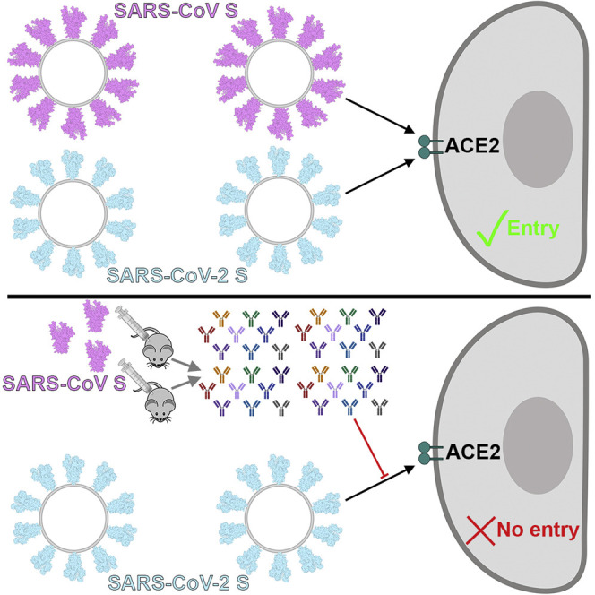
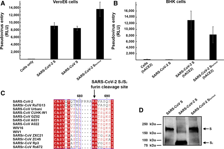
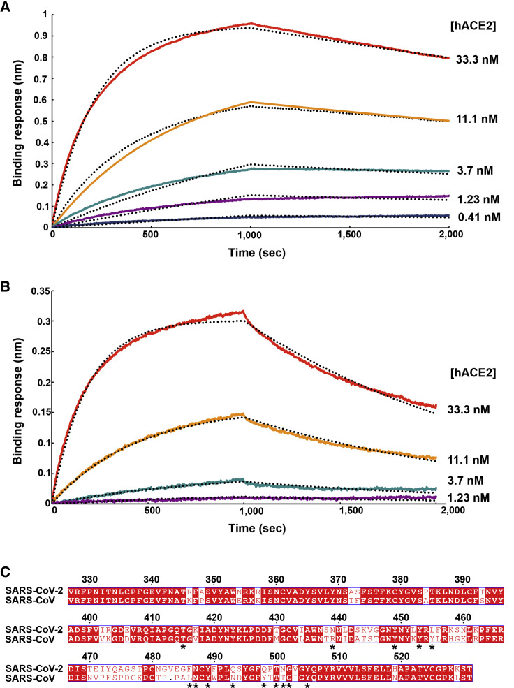
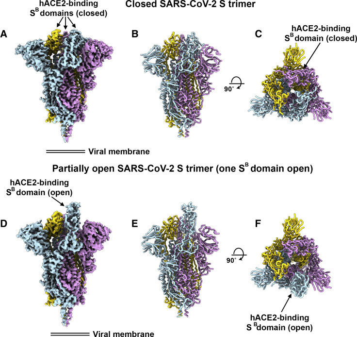
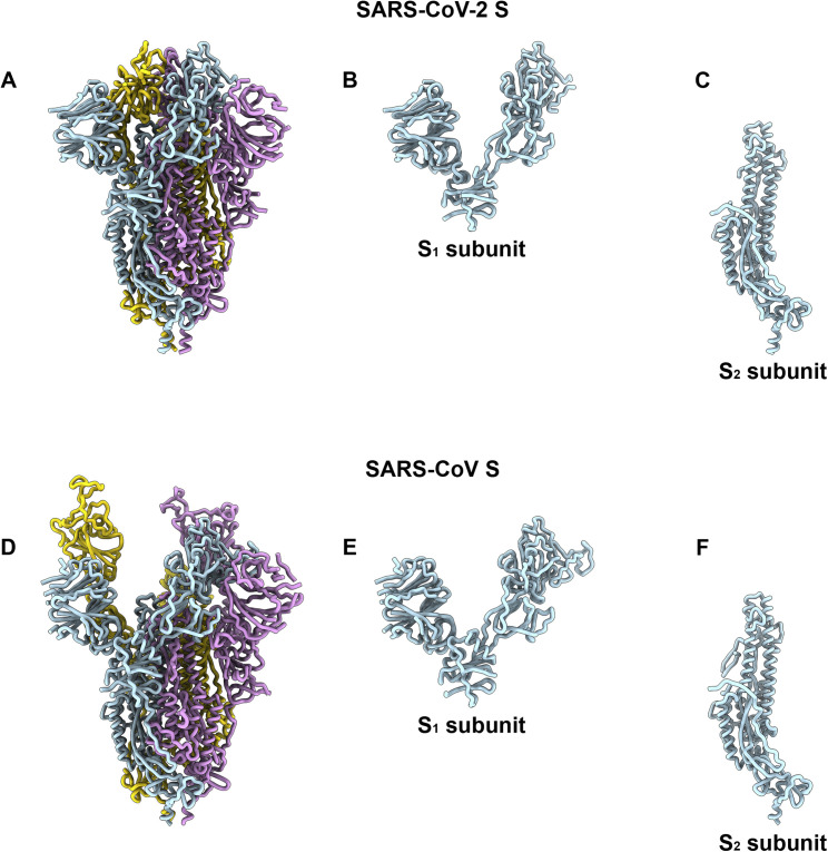
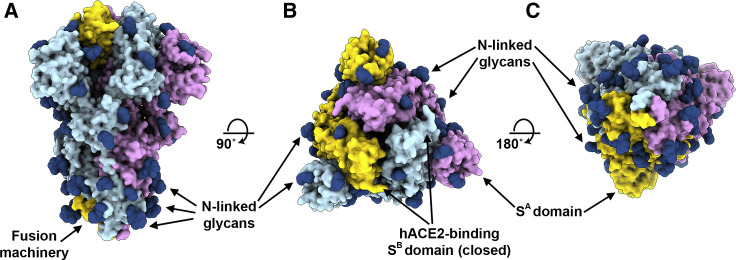
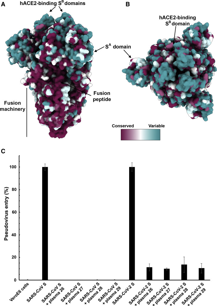
The emergence of SARS-CoV-2 has resulted in >90,000 infections and >3,000 deaths. Coronavirus spike (S) glycoproteins promote entry into cells and are the main target of antibodies. We show that SARS-CoV-2 S uses ACE2 to enter cells and that the receptor-binding domains of SARS-CoV-2 S and SARS-CoV S bind with similar affinities to human ACE2, correlating with the efficient spread of SARS-CoV-2 among humans. We found that the SARS-CoV-2 S glycoprotein harbors a furin cleavage site at the boundary between the S1/S2 subunits, which is processed during biogenesis and sets this virus apart from SARS-CoV and SARS-related CoVs. We determined cryo-EM structures of the SARS-CoV-2 S ectodomain trimer, providing a blueprint for the design of vaccines and inhibitors of viral entry. Finally, we demonstrate that SARS-CoV S murine polyclonal antibodies potently inhibited SARS-CoV-2 S mediated entry into cells, indicating that cross-neutralizing antibodies targeting conserved S epitopes can be elicited upon vaccination.

Keywords: SARS-CoV; SARS-CoV-2; antibodies; coronavirus; cryo-EM; neutralizing antibodies; spike glycoprotein; viral receptor.


Conflict of interest statement

Declaration of Interests The authors declare no competing financial interests.

Figures

None
Graphical abstract
Figure 1
Figure 1
ACE2 Is a Functional Receptor for SARS-CoV-2 S (A) Entry of MLV pseudotyped with SARS-CoV-2 S, SARS-CoV S and SARS-CoV-2 Sfur/mut in VeroE6 cells. Data are represented as mean ± standard deviation of technical triplicates. (B) Entry of MLV pseudotyped with SARS-CoV-2 S or SARS-CoV-2 Sfur/mut in BHK cells transiently transfected with hACE2. The experiments were carried out with two independent pseudovirus preparations and a representative experiment is shown. Data are represented as mean ± standard deviation of technical triplicates. (C) Sequence alignment of SARS-CoV-2 S with multiple related SARS-CoV and SARSr-CoV S glycoproteins reveals the introduction of an S1/S2 furin cleavage site in this novel coronavirus. Identical and similar positions are respectively shown with white or red font. The four amino acid residue insertion at SARS-CoV-2 S positions 681-684 is indicated with periods. The entire sequence alignment is presented in Data S1. (D) Western blot analysis of SARS-CoV S-MLV, SARS-CoV-2 S-MLV, and SARS-CoV-2 Sfur/mut-MLV pseudovirions. See also Data S1.
Figure 2
Figure 2
SARS-CoV-2 S Recognizes hACE2 with Comparable Affinity to SARS-CoV S (A and B) Biolayer interferometry binding analysis of the hACE2 ectodomain to immobilized SARS-CoV-2 SB (A) or SARS-CoV SB (B). The experiments were repeated with different protein preparations and one representative set of curves is shown. Dotted lines correspond to a global fit of the data using a 1:1 binding model. (C) Sequence alignment of SARS-CoV-2 SB and SARS-CoV SB Urbani (late phase of the 2002–2003 SARS-CoV epidemic). Identical and similar positions are respectively shown with white or red font. The single amino acid insertion at position 483 of the SARS-CoV-2 SB is indicated with a period at the corresponding SARS-CoV SB position. The 14 residues that are key for binding of SARS-CoV SB to hACE2 are labeled with a star. See also Data S1.
Figure 3
Figure 3
Cryo-EM Structures of the SARS-CoV-2 S Glycoprotein (A) Closed SARS-CoV-2 S trimer unsharpened cryo-EM map. (B and C) Two orthogonal views from the side (B) and top (C) of the atomic model of the closed SARS-CoV-2 S trimer. (D) Partially open SARS-CoV-2 S trimer unsharpened cryo-EM map (one SB domain is open). (E-F) Two orthogonal views from the side (E) and top (F) of the atomic model of the closed SARS-CoV-2 S trimer. The glycans were omitted for clarity. See also Figures S1 and S2.
Figure S1
Figure S1
Cryo-EM Data Processing and Validation, Related to Figures 3, 4, and 5 A-B. Representative electron micrograph (A) and class averages (B) of SARS-CoV-2 S embedded in vitreous ice. Scale bar: 100nm. C. Gold-standard Fourier shell correlation curves for the closed (blue) and partially open trimers (red). The 0.143 cutoff is indicated by horizontal dashed lines. D-E. Local resolution map calculated using cryoSPARC for the closed (D) and partially open (E) reconstructions.
Figure S2
Figure S2
Comparison of the SARS-CoV-2 and SARS-CoV S Structures, Related to Figures 3, 4, and 5 Ribbon diagrams of the SARS-CoV-2 S (A) and SARS-CoV S (PDB 6NB6, D) ectodomain cryoEM structures. The SARS-CoV-2 (B) and SARS-CoV (E) S1 subunits. The SARS-CoV-2 (C) and SARS-CoV (F) S2 subunits.
Figure 4
Figure 4
Organization of the SARS-CoV-2 S N-Linked Glycans (A–C) Ribbon diagrams of the SARS-CoV-2 S closed structure rendered as a surface with glycans resolved in the cryo-EM map rendered as dark blue spheres. See also Table 2 and Data S1.
Figure 5
Figure 5
SARS-CoV S Elicits Antibodies Neutralizing SARS-CoV-2 S-Mediated Entry into Host Cells (A and B) Sequence conservation of sarbecovirus S glycoproteins plotted on the SARS-CoV-2 S structure viewed from the side (A) and top (B). The sequence alignment was generated using 48 SARS-CoV-2 S sequences obtained from GISAID in addition to the sequences listed in Data S1. (C) Entry of SARS-CoV-2 S-MLV and SARS-CoV S-MLV is potently inhibited by four SARS-CoV S mouse polyclonal immune plasma.

References

    1. Agirre J., Iglesias-Fernández J., Rovira C., Davies G.J., Wilson K.S., Cowtan K.D. Privateer: software for the conformational validation of carbohydrate structures. Nat. Struct. Mol. Biol. 2015;22:833–834. - PubMed
    1. Anthony S.J., Gilardi K., Menachery V.D., Goldstein T., Ssebide B., Mbabazi R., Navarrete-Macias I., Liang E., Wells H., Hicks A. Further Evidence for Bats as the Evolutionary Source of Middle East Respiratory Syndrome Coronavirus. MBio. 2017;8:e00373-17. - PMC - PubMed
    1. Ashkenazy H., Abadi S., Martz E., Chay O., Mayrose I., Pupko T., Ben-Tal N. ConSurf 2016: an improved methodology to estimate and visualize evolutionary conservation in macromolecules. Nucleic Acids Res. 2016;44:W344-50. - PMC - PubMed
    1. Barad B.A., Echols N., Wang R.Y., Cheng Y., DiMaio F., Adams P.D., Fraser J.S. EMRinger: side chain-directed model and map validation for 3D cryo-electron microscopy. Nat. Methods. 2015;12:943–946. - PMC - PubMed
    1. Belouzard S., Chu V.C., Whittaker G.R. Activation of the SARS coronavirus spike protein via sequential proteolytic cleavage at two distinct sites. Proc. Natl. Acad. Sci. USA. 2009;106:5871–5876. - PMC - PubMed

Publication types

MeSH terms