. 2020 Mar 6;10(3):408.
doi: 10.3390/biom10030408.

A Preliminary Study of Cu Exposure Effects upon Alzheimer's Amyloid Pathology

Affiliations

A Preliminary Study of Cu Exposure Effects upon Alzheimer's Amyloid Pathology

Alexander Pilozzi et al. Biomolecules. .

Abstract

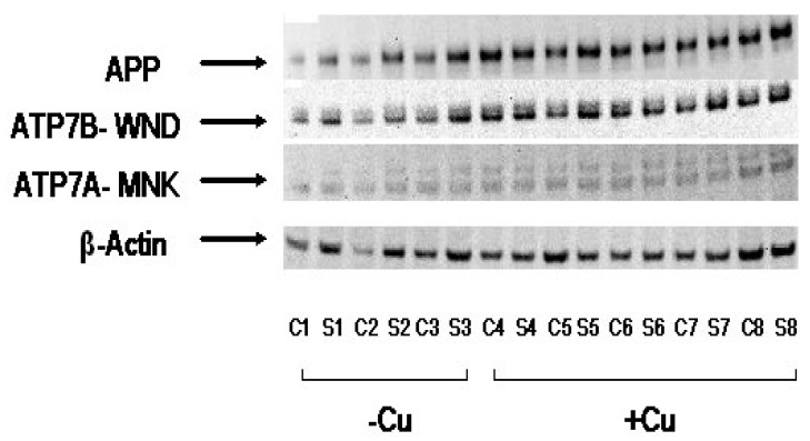
A large body of evidence indicates that dysregulation of cerebral biometals (Fe, Cu, Zn) and their interactions with amyloid precursor protein (APP) and Aβ amyloid may contribute to the Alzheimer's disease (AD) Aβ amyloid pathology. However, the molecular underpinnings associated with the interactions are still not fully understood. Herein we have further validated the exacerbation of Aβ oligomerization by Cu and H2O2 in vitro. We have also reported that Cu enhanced APP translations via its 5' untranslated region (5'UTR) of mRNA in SH-SY5Y cells, and increased Aβ amyloidosis and expression of associated pro-inflammatory cytokines such as MCP-5 in Alzheimer's APP/PS1 doubly transgenic mice. This preliminary study may further unravel the pathogenic role of Cu in Alzheimer's Aβ amyloid pathogenesis, warranting further investigation.

Keywords: Alzheimer’s disease; Aβ amyloid; amyloid precursor protein; copper; cytokine; neuroinflammation.


Conflict of interest statement

The authors declare no conflict of interest.

Figures

Figure 1
Figure 1
Cu and H2O2 exacerbated Aβ oligomerization in vitro. Western blot represented purified Aβ1-40 (11.3 μM) alone (lane 1) or treated with 50 μM Cu2+ (lane 2) or 50 μM Cu2+ + 250 μM H2O2 (lane 3). Bands were detected using an anti-Aβ monoclonal antibody (6E10) that recognizes the N-terminus of Aβ (see Methods for Western blot details).
Figure 2
Figure 2
Cu2+ treatment increased APP protein expression in human SH-SY5Y neuroblastoma cells via 5′UTR of APP mRNA. Human SH-SY5Y neuroblastoma cells stably transfected with APP 5′UTR driven luciferase reporter gene construct with GFP as an internal specificity control- pIRES(APP 5′UTR ) construct, were treated with Cu2+ (0.01, 0.1, 1 µM) for 48 h under regular culture condition (95% O2, 5% CO2, 95% humidity, 37 °C). And then the luciferase substrate (Perkin Elmer, MA, USA) was added and incubated for 30 minutes for complete cell lysis. Luminescence was recorded on an LJL Analyst plate reader (Molecular Device, CA, USA). Percentage activation of luciferase signal was calculated relative to positive and negative controls. The data indicate mean (±SE, n = 5). Inset: Native SH-SY5Y cells were treated with Cu2+ (0, 1 µM) under same culture condition for 3 days. Cells were then washed and lysed, and endogenous expression levels of APP and the control protein- β-actin in lysates were probed by Western blotting.
Figure 3
Figure 3
Dietary Cu exposure enhanced cerebral Aβ amyloid pathology in APP/PS1 transgenic mice. Representative coronal brain sections (at the anterior commissure level) from APP/PS1 transgenic mice on regular and Cu-enhanced diets for 24 days. They were immunostained for Aβ1-42 and Aβ1-40, illustrating the size, number and distribution of Aβ amyloid plaques. The immunostaining for reactive astrogliosis marker- GFAP and negative staining for SYP, a marker for neuronal synaptic activity, were also performed.
Figure 4
Figure 4
Cu treatment increased APP protein expression in APP/PS1 transgenic mouse brain (A) The protein levels of APP, Cu(II)-ATPases- APT7B/WND and ATP7A/MNK, and the control protein- β-actin in lysates were probed by Western blotting. These cortical (C) and striatum (S) tissue lysates were collected from regular food-fed APP/PS1 transgenic mice (3) and mice fed by Cu-enhanced chow (5).
Figure 5
Figure 5
Dietary Cu exposure heightened cerebral pro-inflammatory cytokines in APP/PS1 transgenic mice. 30 different mouse cytokines, chemokines and growth factors in the transgenic mouse cortical lysates were analyzed using Zyomyx Murine Cytokine Biochip System in a multiplexed assay format. Among them, two pro-inflammatory cytokines- MCP-5 and TCA4/6Ckine have shown significant increases in their concentrations due to Cu treatment.

Similar articles

Cited by

References

    1. Alzheimer’s-Association 2019 Alzheimer’s disease facts and figures. Alzheimer’s Dement. 2019;15:321–387. doi: 10.1016/j.jalz.2019.01.010. - DOI - PubMed
    1. Selkoe D.J. Alzheimer’s disease: Genes, proteins, and therapy. Physiol. Rev. 2001;81:741–766. doi: 10.1152/physrev.2001.81.2.741. - DOI - PubMed
    1. Koseoglu E., Koseoglu R., Kendirci M., Saraymen R., Saraymen B. Trace metal concentrations in hair and nails from Alzheimer’s disease patients: Relations with clinical severity. J. Trace Elem. Med. Biol. 2017;39:124–128. doi: 10.1016/j.jtemb.2016.09.002. - DOI - PubMed
    1. Squitti R., Ghidoni R., Simonelli I., Ivanova I.D., Colabufo N.A., Zuin M., Benussi L., Binetti G., Cassetta E., Rongioletti M., et al. Copper dyshomeostasis in Wilson disease and Alzheimer’s disease as shown by serum and urine copper indicators. J. Trace Elem. Med. Biol. 2018;45:181–188. doi: 10.1016/j.jtemb.2017.11.005. - DOI - PubMed
    1. Vaz F.N.C., Fermino B.L., Haskel M.V.L., Wouk J., de Freitas G.B.L., Fabbri R., Montagna E., Rocha J.B.T., Bonini J.S. The relationship between copper, iron, and selenium levels and alzheimer disease. Biol. Trace Elem. Res. 2018;181:185–191. doi: 10.1007/s12011-017-1042-y. - DOI - PubMed

Publication types