. 2020 Mar 6;10(3):412.
doi: 10.3390/biom10030412.

Origanum majorana Essential Oil Triggers p38 MAPK-Mediated Protective Autophagy, Apoptosis, and Caspase-Dependent Cleavage of P70S6K in Colorectal Cancer Cells

Affiliations

Origanum majorana Essential Oil Triggers p38 MAPK-Mediated Protective Autophagy, Apoptosis, and Caspase-Dependent Cleavage of P70S6K in Colorectal Cancer Cells

Khawlah Athamneh et al. Biomolecules. .

Abstract

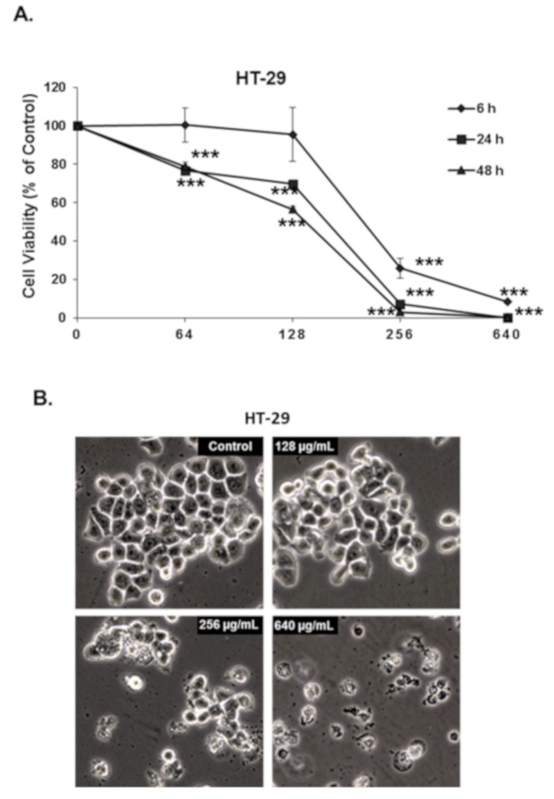
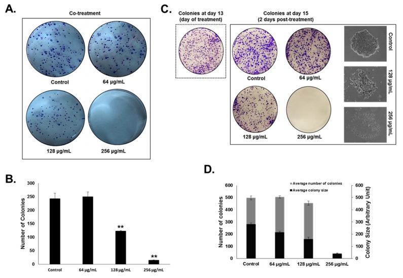
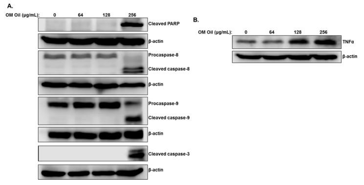
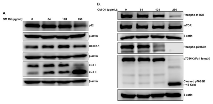
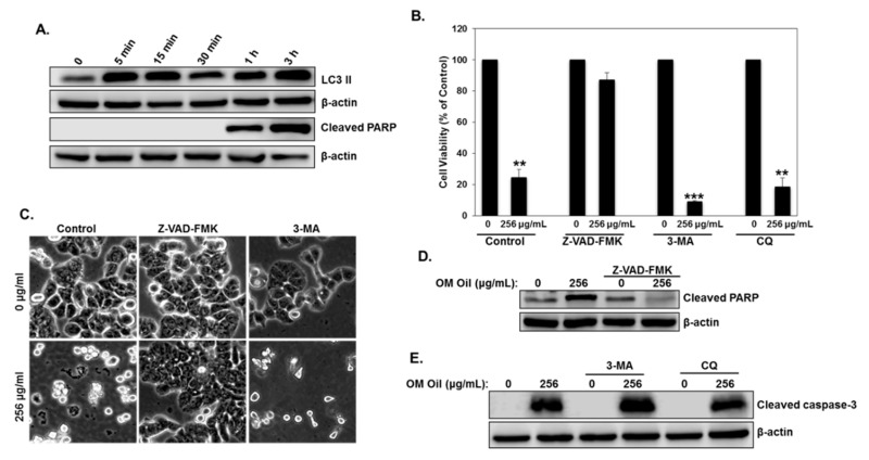
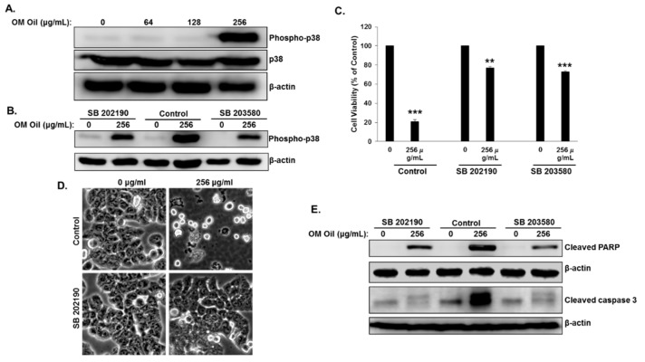
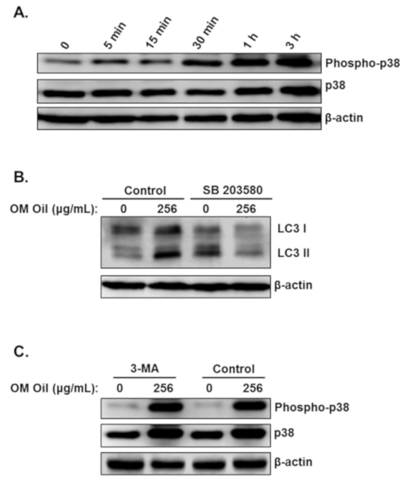
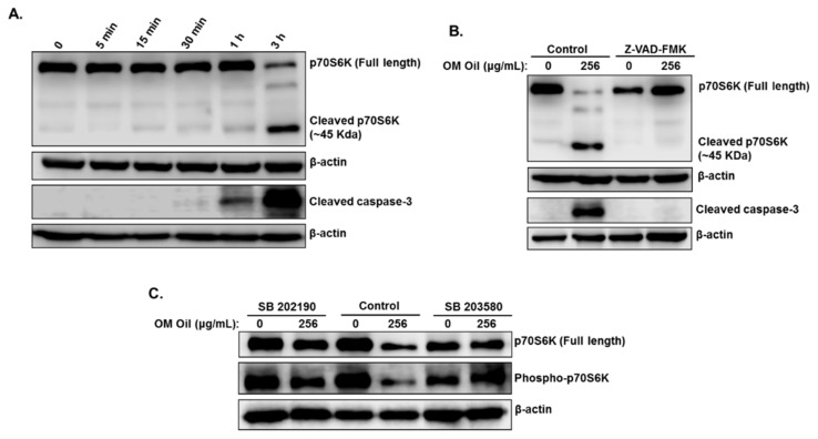
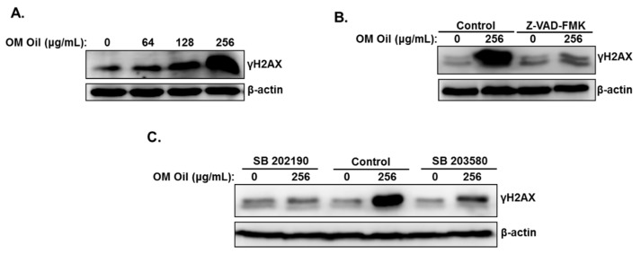
Colorectal cancer (CRC) is the third most common type of cancer in terms of incidence and mortality worldwide. Here we have investigated the anti-colon cancer potential of Origanum majorana essential oil (OMEO) and its underlying mechanisms of action. We showed that OMEO significantly inhibited the cellular viability and colony growth of human HT-29 colorectal cancer cells. OMEO induced protective autophagy, associated with downregulation of the mTOR/p70S6K pathway, and activated caspase-8 and caspase-9-dependent apoptosis. Blockade of autophagy with 3-methyladenine (3-MA) and chloroquine (CQ), two autophagy inhibitors, potentiated the OMEO-induced apoptotic cell death. Inversely, inhibition of apoptosis with the pan-caspase inhibitor, Z-VAD-FMK, significantly reduced cell death, suggesting that apoptosis represents the main mechanism of OMEO-induced cell death. Mechanistically, we found that OMEO induces protective autophagy and apoptotic cells death via the activation of the p38 MAPK signaling pathway. Pharmacological inhibition of p38 MAPK by the p38 inhibitors SB 202190 and SB 203580 not only significantly decreased apoptotic cell death, but also reduced the autophagy level in OMEO treated HT-29 cells. Strikingly, we found that OMEO also induces p38 MAPK-mediated caspase-dependent cleavage of p70S6K, a protein reported to be overexpressed in colon cancer and associated with drug resistance. Our findings suggest that OMEO inhibits colon cancer through p38 MAPK-mediated protective autophagy and apoptosis associated with caspase-dependent cleavage of p70S6K. To the best of our knowledge, this study is the first to report on the implications of the p38 MAPK signaling pathway in targeting p70S6K to caspase cleavage.

Keywords: Origanum majorana; apoptosis; autophagy; colon cancer; p38MAPK; p70S6K..


Conflict of interest statement

The authors declare that the research was conducted in the absence of any commercial or financial relationships that could be construed as a potential conflict of interest.

Figures

Figure 1
Figure 1
Origanum majorana essential oil (OMEO) inhibits the cellular viability of HT-29 cells. (A) Exponentially growing HT-29 colon cancer cells were treated with and without the indicated concentrations (0, 64, 128, 256, and 640 μg/mL) of OMEO for 6, 24, and 48 h. Viability was measured as described in Section 2.3 Data represent the mean of six independent experiments carried out in triplicate. Values are represented as mean ± SD of n = 4 (*** p < 0.001). (B) Morphological changes in OMEO-treated HT-29 cells. Morphological changes observed in the treated HT-29 cells after 6 h of treatment with the indicated concentration of OMEO. Cells were observed under EVOS XL Core Cell Imaging System (Life Technologies) at 400×.
Figure 2
Figure 2
OMEO inhibits HT-29 colony growth. (A,B) Inhibition of HT-29 colony growth by various concentrations of OMEO (0, 64, 128, and 256 μg/mL) was assessed by measuring the number of the colonies obtained in control and OMEO-treated plate, as described in Section 2.5 Values are represented as mean ± SD of n = 3 (** p < 0.005). (C) HT-29 colonies were first allowed to form in normal media for 13 days as described in Section 2.5 Formed colonies were then treated with or without increasing concentrations of OMEO, and allowed to grow for three more days before crystal violet staining. The size and morphology of the growing colonies was tracked over time with the EVOS XL Core Cell Imaging System (Life Technologies) at 400×. (D) Inhibition of colony growth was assessed by measuring the number and size (surface area) of the colonies obtained in control and OMEO-treated plates, as described in Section 2.5 Data represent the mean of three independent experiments carried out in duplicate. Values are represented as mean ± SD of n = 3 (* p < 0.05, ** p < 0.005).
Figure 3
Figure 3
Induction of caspase-8, -9, and -3-mediated apoptosis by OMEO in HT-29 cells. (A) Western blot analysis of cleaved PARP, caspase-8, -9, and -3 activation in HT-29 cells treated with increasing concentrations of OMEO (0, 64, 128, and 256 μg/mL) for 6 h. (B) Western blot quantification of TNF-α protein in OMEO-treated HT-29 cells.
Figure 4
Figure 4
OMEO induces autophagy associated with downregulation of mTOR/p70S6K in HT-29 cells. (A) Induction of autophagy by OMEO. Western blotting analysis of marker of autophagy, p62(SQSTM1), Beclin-1, and LC3 II in OMEO-treated HT-29 cells. Cells were treated with or without an increasing concentration of OMEO (0, 64, 128, and 256 μg/mL) for 6 h, then whole cell proteins were extracted and subjected to Western blot analysis, as described in Section 2.6 (B) Downregulation of the mTOR/p70S6K by OMEO. Western blot analysis for the phosphorylated and non-phosphorylated form of mTOR and p70S6K.
Figure 5
Figure 5
Protective autophagy and apoptotic cell death in HT-29 cells in response to OMEO. (A) Time-course analysis of LC3 II and cleaved PARP in OMEO-treated HT-29 cells. Cells were treated with 256 μg/mL OMEO and protein levels were determined by Western blot at different time points (0, 5 min, 15 min, 30 min, 1 h, and 3 h) post-treatment. (B) Blockade of autophagy increases cell death, while inhibition of apoptosis promotes cell survival. HT-29 cells were pretreated with Z-VAD-FMK, 3-MA, or CQ and then incubated for 6 h with 256 μg/mL OMEO. Cell viability was determined as described in Section 2.3 (C) Micrograph observation of HT-29 cells pretreated with or without pan-caspase inhibitor (Z-VAD-FMK) and 3-MA, as described above. (D) Western blotting analysis of cleaved PARP in OMEO cell pretreated with Z-VAD-FMK. (E) Western blotting analysis of cleaved caspase-3 with or without 3-MA or CQ.
Figure 6
Figure 6
p38 MAPK-dependent activation of apoptosis in OMEO-treated cells. (A) OMEO activates p38 MAPK in HT-29 cells. Western blotting analysis of phosphorylated and total p38 in OMEO-treated HT-29 cells. Cells were treated with or without an increasing concentration (0, 64, 128, and 256 μg/mL) for 6 h, then whole cell proteins were subjected to Western blot analysis. (B) Western blotting analysis of phosphorylated p38 in the presence of p38 inhibitors (SB 202190 and SB 203580). (C) Inhibition of p38 MAPK abrogates the OMEO-induced apoptotic cell death. HT-29 cells were pretreated with p38 inhibitors and cellular viability was determined as described in Section 2.3 (D) Morphological changes observed in the treated HT-29 pretreated with p38 inhibitors prior the incubation with OMEO. Cells were observed under EVOS XL Core Cell Imaging System (Life Technologies) at 400×. (E) Western blotting analysis of cleaved PARP and cleaved caspase-3 in cell pretreated with and without SB 202190 and SB 203580.
Figure 7
Figure 7
p38 MAPK-mediated protective autophagy. (A) Time-course measurement of phosphorylated and total p38 level in OMEO-treated cells. Cells were treated with 256 μg/mL OMEO and the level of active and total p38 was examined at different time points (0, 5 min, 15 min, 30 min, 1 h and 3 h). (B) LC3 I and LC3 II levels were measured in cells pretreated with SB 203580. (C) Western blotting analysis of phosphorylated and total p38 in cells pretreated with and without 3-MA.
Figure 8
Figure 8
p38 MAPK-dependent caspase-dependent cleavage of p70S6K. (A) Time course measurement of p70S6K and cleaved caspase-3 in OMEO-treated HT-29 cells. Cells were treated with 256 μg/mL OMEO and protein levels were examined at different time points (0, 5 min, 15 min, 30 min, 1 h, and 3 h). (B) Western blotting analysis of p70S6K and cleaved caspase-3 in cells pretreated with Z-VAD-FMK. (C) Western blot analysis of full-length and phosphorylated p70S6K in cells pretreated with SB 202190 and SB 203580.
Figure 9
Figure 9
DNA damage in OMEO-treated HT-29 cells. (A) Accumulation of γH2AX, a marker of DNA damage, in OMEO-treated cells. HT-29 cells were treated with and without increasing concentrations of OMEO for 6 h and DNA damage was analyzed by Western blot, by determining the level of γH2AX accumulation. (B) Western blotting analysis of γH2AX in cells pretreated with and without Z-VAD-FMK. (C) Western blotting analysis of γH2AX in cells pretreated with and without SB 202190 and SB 203580.

References

    1. Kuipers E.J., Grady W.M., Lieberman D., Seufferlein T., Sung J.J., Boelens P.G., van de Velde C.J.H., Watanabe T. Colorectal cancer. Nat. Rev. Dis. Primers. 2015;1:15065. doi: 10.1038/nrdp.2015.65. - DOI - PMC - PubMed
    1. Peluso G., Incollingo P., Calogero A., Tammaro V., Rupealta N., Chiacchio G., Sandoval Sotelo M.L., Minieri G., Pisani A., Riccio E., et al. Current Tissue Molecular Markers in Colorectal Cancer: A Literature Review. Biomed. Res. Int. 2017;2017:2605628. doi: 10.1155/2017/2605628. - DOI - PMC - PubMed
    1. Newman D.J., Cragg G.M. Natural Products as Sources of New Drugs from 1981 to 2014. J. Nat. Prod. 2016;79:629–661. doi: 10.1021/acs.jnatprod.5b01055. - DOI - PubMed
    1. Balunas M.J., Kinghorn A.D. Drug discovery from medicinal plants. Life Sci. 2005;78:431–441. doi: 10.1016/j.lfs.2005.09.012. - DOI - PubMed
    1. Nobili S., Lippi D., Witort E., Donnini M., Bausi L., Mini E., Capaccioli S. Natural compounds for cancer treatment and prevention. Pharmacol. Res. Off. J. Ital. Pharmacol. Soc. 2009;59:365–378. doi: 10.1016/j.phrs.2009.01.017. - DOI - PubMed

Publication types

MeSH terms