. 2020 Mar 6;9(3):645.
doi: 10.3390/cells9030645.

MicroRNAs in Small Extracellular Vesicles Indicate Successful Embryo Implantation during Early Pregnancy

Affiliations

MicroRNAs in Small Extracellular Vesicles Indicate Successful Embryo Implantation during Early Pregnancy

Qiang Tan et al. Cells. .

Abstract

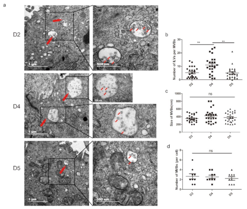
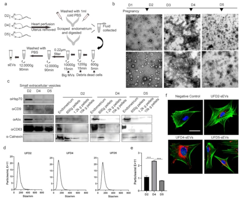
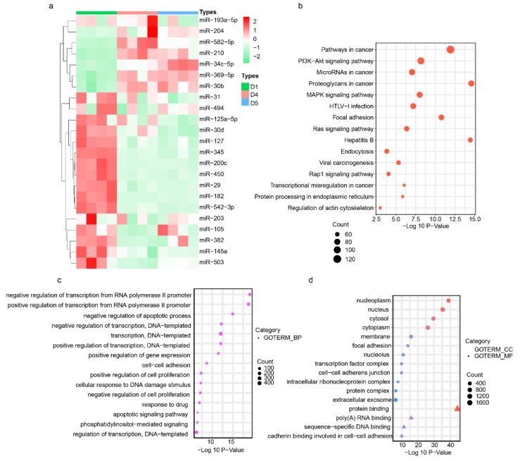
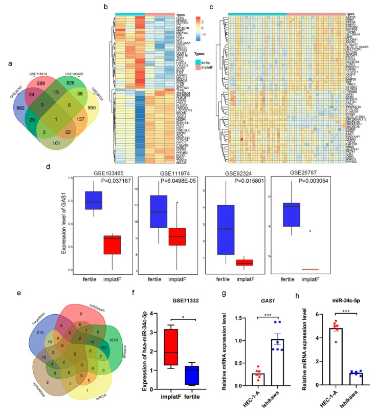
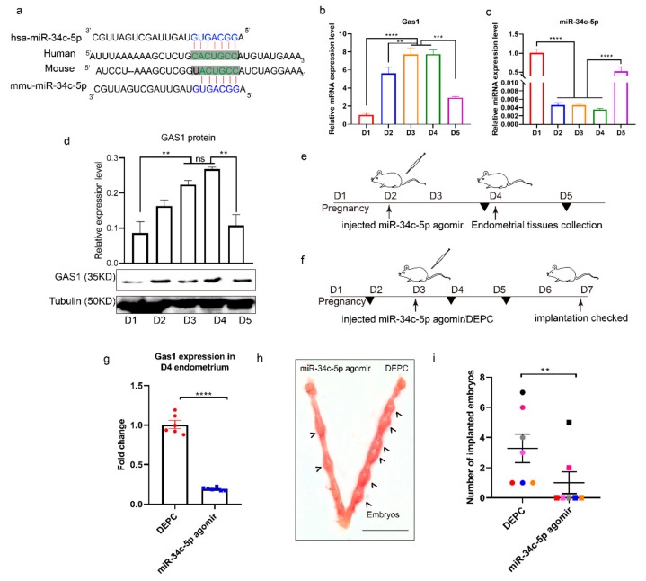
Synchronous communication between the developing embryo and the receptive endometrium is crucial for embryo implantation. Thus, uterine receptivity evaluation is vital in managing recurrent implantation failure (RIF). The potential roles of small extracellular vesicle (sEV) miRNAs in pregnancy have been widely studied. However, the systematic study of sEVs derived from endometrium and its cargos during the implantation stage have not yet been reported. In this study, we isolated endometrium-derived sEVs from the mouse endometrium on D2 (pre-receptive phase), D4 (receptive phase), and D5 (implantation) of pregnancy. Herein, we reveal that multivesicular bodies (MVBs) in the endometrium increase in number during the window of implantation (WOI). Moreover, our findings indicate that CD63, a well-known sEV marker, is expressed in the luminal and glandular epithelium of mouse endometrium. The sEV miRNA expression profiles indicated that miR-34c-5p, miR-210, miR-369-5p, miR-30b, and miR-582-5p are enriched during WOI. Further, we integrated the RIF's database analysis results and found out that miR-34c-5p regulates growth arrest specific 1 (GAS1) for normal embryo implantation. Notably, miR-34c-5p is downregulated during implantation but upregulated in sEVs. An implication of this is the possibility that sEVs miR-34c-5p could be used to evaluate uterine states. In conclusion, these findings suggest that the endometrium derived-sEV miRNAs are potential biomarkers in determining the appropriate period for embryo implantation. This study also has several important implications for future practice, including therapy of infertility.

Keywords: embryo implantation; miRNAs; recurrent implantation failure; small extracellular vesicles.


Conflict of interest statement

The authors declare no conflict of interest.

Figures

Figure 1
Figure 1
MVBs are present in mouse endometrium cells during early pregnancy. (a) Representative electron microscopic images of endometrium tissues on D2, D4, and D5 of pregnancy: Red arrows indicate MVBs including classic intraluminal vesicles (ILVs). (b) The number of ILVs per MVB, (c) size of MVBs, (d) number of MVBs per cell in different stages: MVBs were counted only when containing typical ILVs. ** P < 0.01. ns: not significant.
Figure 2
Figure 2
Immunohistochemical staining of CD63 in mouse glandular and luminal epithelium during pre-implantation (D2), window of implantation (WOI) (D4), post-implantation (D5) stage, and in the negative control (no primary antibody). LE: luminal epithelium. GE: glandular epithelium. IS: implantation site. IIS: inter implantation site.
Figure 3
Figure 3
Identification of small extracellular vesicles (sEVs) derived from endometrium: (a) Schematic diagram showed the strategy of isolation and purification of sEVs derived from mouse endometrium during early pregnancy. (b) TEM images showed sEVs derived from mouse endometrium on D2, D4, and D5 of pregnancy. Magnification: top, 18,500. bottom, 68,000. (c) Western blotting analysis of sEV protein markers (CD63, Alix, CD9, and HSP70): Calnexin was used as a negative control. (d) Nanoparticle Tracking Analysis (NTA)-suggested size distribution and concentration of sEVs. (e) Concentration of sEVs derived from the uterus on D2, D4, and D5 of pregnancy. (f) Confocal microscopic images showed uptake of labeled sEVs by primary endometrium cells (pECs). Nuclei are stained by DAPI in blue, sEVs are stained by DiI in red, and actin is stained by FITC-conjugated phalloidin in green. (Bar 20 μm). *** P < 0.001.
Figure 4
Figure 4
Endometrium-derived sEVs carry miRNAs which associated with uterine receptivity: (a) Heat map showed the expression profiles of miRNAs in sEVs derived from endometrium on D2, D4, and D5 of pregnancy. (bd) GeneOntology GeneOntology (GO) and Kyoto Encyclopedia of Genes and Genomes (KEGG)terms of genes were potentially targeted by miRNAs in sEVs. (b) A bubble plot of KEGG pathways enriched. (c) A bubble plot of GOs (Biological Process) enriched. (d) A bubble plot of GOs (molecular function and cellular components) enriched.
Figure 5
Figure 5
Growth arrest specific 1 (GAS1) was targeted by miR-34c-5p. (a) The Venn diagram of co-expressed differentially expressed genes (DEGs) in four Gene Expression Omnibus (GEO) databases (GSE26787, GSE111974, GSE103465, and GSE92324): only one gene, GAS1, was observed. (b,c) Heat maps show the top 60 differentially expressed genes in GSE103465 and GSE111974, respectively. (d) The expression of GAS1 in four gene microarrays. (e) The comparison of target miRNA of GAS1 predicted by TargetScan, miRSearch, miRTarBase, miRWalk, and mirDIP. (f) The expression of hsa-miR-34c-5p in miRNA expression chip GEO: GSE71332 of RIF. (g) The mRNA level of GAS1 in Ishikawa and HEC-1-A cell lines. (h) The expression of miR-34c-5p in Ishikawa and HEC-1-A cell lines. * P < 0.05, *** P < 0.001.
Figure 6
Figure 6
miR-34c-5p regulated embryo implantation by targeting GAS1 in endometrium during early pregnancy. (a) The regulating relationship between GAS1 and miR-34c-5p, the mature sequence of miR-34c-5p in mouse and human, was predicted by Targetscan. (b,c) The relative expression of GAS1 and miR-34c-5p in mouse endometrium was detected by qRT-PCR during early pregnancy. (d) The protein expression level of GAS1 in mouse endometrium. (e) miR-34c-5p regulated the expression of GAS1 in endometrium. (f) miR-34c-5p regulated successful embryo implantation. (g) The expression of GAS1 in D4 mouse endometrium after injecting miR-34c-5p agomir in D2. (h,i) The effect of miR-34c-5p agomir injected in vivo on the number of embryos. Bar = 1 cm. ** P < 0.01, *** P < 0.001.

References

    1. Bashiri A., Halper K.I., Orvieto R. Recurrent Implantation Failure-update overview on etiology, diagnosis, treatment and future directions. Reprod. Biol. Endocrinol. 2018;16:121. doi: 10.1186/s12958-018-0414-2. - DOI - PMC - PubMed
    1. Coughlan C., Ledger W., Wang Q., Liu F., Demirol A., Gurgan T., Cutting R., Ong K., Sallam H., Li T.C. Recurrent implantation failure: Definition and management. Reprod. Biomed. Online. 2014;28:14–38. doi: 10.1016/j.rbmo.2013.08.011. - DOI - PubMed
    1. Wilcox A.J., Baird D.D., Weinberg C.R. Time of implantation of the conceptus and loss of pregnancy. N. Engl. J. Med. 1999;340:1796–1799. doi: 10.1056/NEJM199906103402304. - DOI - PubMed
    1. Sharma A., Kumar P. Understanding implantation window, a crucial phenomenon. J. Hum. Reprod. Sci. 2012;5:2–6. - PMC - PubMed
    1. Wang H., Dey S.K. Roadmap to embryo implantation: Clues from mouse models. Nat. Rev. Genet. 2006;7:185–199. doi: 10.1038/nrg1808. - DOI - PubMed

Publication types

LinkOut - more resources