Diagnostic Performance of a Combination of Shear Wave Elastography and B-Mode Ultrasonography in Differentiating Benign From Malignant Thyroid Nodules
- PMID: 32156104
- PMCID: PMC7248619
- DOI: 10.21053/ceo.2019.01235
Diagnostic Performance of a Combination of Shear Wave Elastography and B-Mode Ultrasonography in Differentiating Benign From Malignant Thyroid Nodules
Abstract
Objectives: This study was conducted to compare clinicopathologic and radiologic factors between benign and malignant thyroid nodules and to evaluate the diagnostic performance of shear wave elastography (SWE) combined with B-mode ultrasonography (US) in differentiating malignant from benign thyroid nodules.
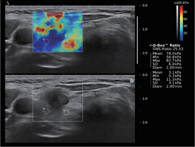
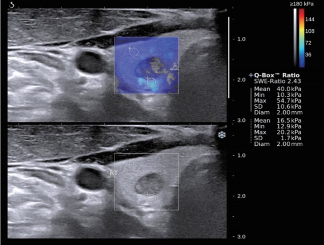
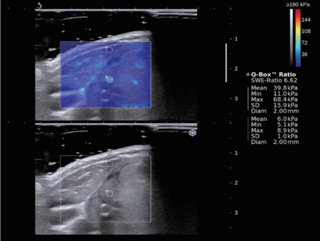
Methods: This retrospective study included 92 consecutive patients with 95 thyroid nodules examined on B-mode US and SWE before US-guided fine-needle aspiration biopsy or surgical excision. B-mode US findings (composition, echogenicity, margin, shape, and calcification) and SWE elasticity parameters (maximum [Emax], mean, minimum, and nodule-to-normal parenchymal ratio of elasticity) were reviewed and compared between benign and malignant thyroid nodules. The diagnostic performance of B-mode US and SWE for predicting malignant thyroid nodules was analyzed. The optimal cutoff values of elasticity parameters for identifying malignancy were determined. Diagnostic performance was compared between B-mode US only, SWE only, and the combination of B-mode US with SWE.
Results: On multivariate logistic regression analysis, age (odds ratio [OR], 0.90; P=0.028), a taller-than-wide shape (OR, 11.3; P=0.040), the presence of calcifications (OR, 15.0; P=0.021), and Emax (OR, 1.22; P=0.021) were independent predictors of malignancy in thyroid nodules. The combined use of B-mode US findings and SWE yielded improvements in sensitivity, the positive predictive value, the negative predictive value, and accuracy compared with the use of B-mode US findings only, but with no statistical significance.
Conclusion: When SWE was combined with B-mode US, the diagnostic performance was better than when only B-mode US was used, although the difference was not statistically significant.
Keywords: Clinicopathologic and Radiologic Factors; Diagnostic Performance; Shear Wave Elastography; Thyroid; Ultrasound.
Conflict of interest statement
No potential conflict of interest relevant to this article was reported.
Figures
References
-
- Kim EK, Park CS, Chung WY, Oh KK, Kim DI, Lee JT, et al. New sonographic criteria for recommending fine-needle aspiration biopsy of nonpalpable solid nodules of the thyroid. AJR Am J Roentgenol. 2002 Mar;178(3):687–91. - PubMed
-
- Hoang JK, Lee WK, Lee M, Johnson D, Farrell S. US Features of thyroid malignancy: pearls and pitfalls. Radiographics. 2007 May-Jun;27(3):847–60. - PubMed
-
- Iannuccilli JD, Cronan JJ, Monchik JM. Risk for malignancy of thyroid nodules as assessed by sonographic criteria: the need for biopsy. J Ultrasound Med. 2004 Nov;23(11):1455–64. - PubMed
-
- Alexander EK. Approach to the patient with a cytologically indeterminate thyroid nodule. J Clin Endocrinol Metab. 2008 Nov;93(11):4175–82. - PubMed