Neural stem cell-conditioned medium ameliorates Aβ25-35-induced damage in SH-SY5Y cells by protecting mitochondrial function
- PMID: 32156251
- PMCID: PMC7982066
- DOI: 10.17305/bjbms.2020.4570
Neural stem cell-conditioned medium ameliorates Aβ25-35-induced damage in SH-SY5Y cells by protecting mitochondrial function
Abstract
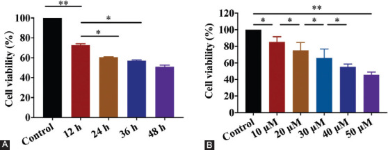
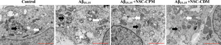
Inhibition of amyloid β (Aβ)-induced mitochondrial damage is considered crucial for reducing the pathological damage in Alzheimer's disease (AD). We evaluated the effect of neural stem cell-conditioned medium (NSC-CDM) on Aβ25-35-induced damage in SH-SY5Y cells. An in vitro model of AD was established by treating SH-SY5Y cells with 40 µM Aβ25-35 for 24 h. SH-SY5Y cells were divided into control, Aβ25-35 (40 µM), Aβ25-35 (40 µM) + NSC-CDM, and Aβ25-35 (40 µM) + neural stem cell-complete medium (NSC-CPM) groups. Cell viability was detected by CCK-8 assay. Apoptosis, reactive oxygen species (ROS) production, and mitochondrial membrane potential (MMP) were detected by flow cytometry. Malondialdehyde content was detected by ELISA assay. Western blot analysis was used to detect cytochrome c release and apoptosis-related proteins. Transmission electron microscopy was used to observe mitochondrial morphology. Cell viability significantly decreased and apoptosis significantly increased in SH-SY5Y cells treated with Aβ25-35, and both effects were rescued by NSC-CDM. In addition, NSC-CDM reduced ROS production and significantly inhibited the reduction of MMP caused by Aβ25-35. Furthermore, NSC-CDM ameliorated Aβ25-35-induced reduction in Bcl-2 expression levels and increased the expression levels of cytochrome c, caspase-9, caspase-3, and Bax. Moreover, Aβ25-35 induced the destruction of mitochondrial ultrastructure and this effect was reversed by NSC-CDM. Collectively, our findings demonstrated the protective effect of NCS-CDM against Aβ25-35-induced SH-SY5Y cell damage and clarified the mechanism of action of Aβ25-35 in terms of mitochondrial maintenance and mitochondria-associated apoptosis signaling pathways, thus providing a theoretical basis for the development of novel anti-AD treatments.
Conflict of interest statement
Conflict of interest statement: The authors declare no conflict of interests
Figures


Similar articles
-
Neural stem cell conditioned medium alleviates Aβ25-35 damage to SH-SY5Y cells through the PCMT1/MST1 pathway.Eur J Histochem. 2020 Jun 19;64(s2):3135. doi: 10.4081/ejh.2020.3135. Eur J Histochem. 2020. Retraction in: Eur J Histochem. 2020 Oct 16;64(s2). doi: 10.4081/ejh.2020.3190. PMID: 32705859 Free PMC article. Retracted.
-
Olfactory Ensheathing Cell-Conditioned Medium Reverts Aβ25-35-Induced Oxidative Damage in SH-SY5Y Cells by Modulating the Mitochondria-Mediated Apoptotic Pathway.Cell Mol Neurobiol. 2017 Aug;37(6):1043-1054. doi: 10.1007/s10571-016-0437-1. Epub 2016 Nov 2. Cell Mol Neurobiol. 2017. PMID: 27807758 Free PMC article.
-
Protective Effect of Edaravone Against Aβ25-35-Induced Mitochondrial Oxidative Damage in SH-SY5Y Cells.Cell Mol Biol (Noisy-le-grand). 2017 May 20;63(5):36-42. doi: 10.14715/cmb/2017.63.5.8. Cell Mol Biol (Noisy-le-grand). 2017. PMID: 28719344
-
SOD3 Ameliorates Aβ25-35-Induced Oxidative Damage in SH-SY5Y Cells by Inhibiting the Mitochondrial Pathway.Cell Mol Neurobiol. 2017 Apr;37(3):513-525. doi: 10.1007/s10571-016-0390-z. Epub 2016 Jun 7. Cell Mol Neurobiol. 2017. PMID: 27272114 Free PMC article.
-
Inhibition of beta-amyloid-induced neurotoxicity by pinocembrin through Nrf2/HO-1 pathway in SH-SY5Y cells.J Neurol Sci. 2016 Sep 15;368:223-30. doi: 10.1016/j.jns.2016.07.010. Epub 2016 Jul 11. J Neurol Sci. 2016. PMID: 27538638
Cited by
-
Human neural stem cell secretome relieves endoplasmic reticulum stress-induced apoptosis and improves neuronal functions after traumatic brain injury in a rat model.J Mol Histol. 2024 Jun;55(3):329-348. doi: 10.1007/s10735-024-10192-7. Epub 2024 Apr 12. J Mol Histol. 2024. PMID: 38609527
-
Oxidative-Signaling in Neural Stem Cell-Mediated Plasticity: Implications for Neurodegenerative Diseases.Antioxidants (Basel). 2021 Jul 6;10(7):1088. doi: 10.3390/antiox10071088. Antioxidants (Basel). 2021. PMID: 34356321 Free PMC article. Review.
References
-
- Shah H, Albanese E, Duggan C, Rudan I, Langa KM, Carrillo MC, et al. Research priorities to reduce the global burden of dementia by 2025. Lancet Neurol. 2016;15(12):1285–94. https://doi.org/10.1016/s1474-4422(16)30235-6. - PubMed
-
- Forner S, Baglietto-Vargas D, Martini AC, Trujillo-Estrada L, LaFerla FM. Synaptic impairment in Alzheimer's disease:A dysregulated symphony. Trends Neurosci. 2017;40(6):347–57. https://doi.org/10.1016/j.tins.2017.04.002. - PubMed
-
- Leuner K, Müller WE, Reichert AS. From mitochondrial dysfunction to amyloid beta formation:Novel insights into the pathogenesis of Alzheimer's disease. Mol Neurobiol. 2012;46(1):186–93. https://doi.org/10.1007/s12035-012-8307-4. - PubMed
-
- Rubio-Moscardo F, Setó-Salvia N, Pera M, Bosch-Morató M, Plata C, Belbin O, et al. Rare variants in calcium homeostasis modulator 1 (CALHM1) found in early onset Alzheimer's disease patients alter calcium homeostasis. PLoS One. 2013;8(9):e74203. https://doi.org/10.1371/journal.pone.0074203. - PMC - PubMed
-
- Landgren H, Curtis MA. Locating and labeling neural stem cells in the brain. J Cell Physiol. 2011;226(1):1–7. https://doi.org/10.1002/jcp.22319. - PubMed
MeSH terms
Substances
LinkOut - more resources
Full Text Sources
Research Materials
Miscellaneous