

Global diastolic strain rate for the assessment of left ventricular diastolic dysfunction in young peritoneal dialysis patients: a case control study
- PMID: 32156262
- PMCID: PMC7063726
- DOI: 10.1186/s12882-020-01742-8
Global diastolic strain rate for the assessment of left ventricular diastolic dysfunction in young peritoneal dialysis patients: a case control study
Abstract
Background: Left ventricular (LV) myocardial longitudinal diastolic strain rate measured by two-dimensional speckle tracking imaging (2D-STI) was proved to have a better correlation with the LV diastolic function. We aimed to use this sensitive tool to predict LV myocardial diastolic dysfunction in young peritoneal dialysis (PD) patients with preserved LV ejection fraction (LVEF).
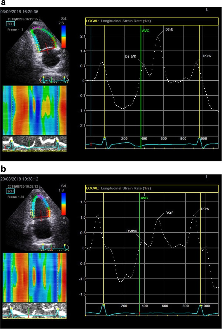
Methods: We enrolled 30 PD patients aged ≤60 with LVEF ≥54% and classified as normal LV diastolic function by conventional echocardiography, and 30 age- and sex-matched healthy people as the control group. The left atrial maximum volume index (LAVI), LV mass index (LVMI), LVEF, LV posterior wall thickness (LVPWT), interventricular septal thickness (IVST), peak velocity of tricuspid regurgitation (TR), peak early diastolic velocity/late diastolic velocity (by Pulsed Doppler) (E/A) and E/peak velocity of the early diastolic wave (by Pulsed-wave tissue Doppler) (E/e') were recorded by conventional echocardiographic. Next, the average LV global longitudinal systolic strain (GLS avg) and the average LV global longitudinal diastolic strain rate (DSr avg) during early diastole (DSrE avg), late diastole (DSrA avg) and isovolumic relaxation period (DSrIVR avg) were obtained from 2D-STI. Combined them with E, the new noninvasive indexes (E/DSrE avg., E/DSrA avg. and E/DSrIVR avg) were derived.
Results: The PD group 's LVEF, E/e', TR and LAVI were in the normal range compared with the controls, and only e' (p < 0.001) was decreased. The LVMI (p < 0.001), LVPWT (p < 0.001), IVST (p < 0.001) increased while E/A (p < 0.001) decreased. The GLS avg. (p = 0.008) was significantly decreased in PD patients compared with the controls. DSrA avg. (p = 0.006) and E/DSrE avg. (p = 0.006) were increased, while DSrE avg. (p < 0.001), DSrIVR avg. (p = 0.017) and E/DSrA avg. (p < 0.001) decreased. After the multivariable regression analysis, the correlation between DSrE and the conventional parameters including LVPWT (p < 0.001), E/A (p < 0.001) still remained significant.
Conclusions: Young PD patients with preserved LVEF already exhibited myocardial diastolic dysfunction. Global diastolic strain rate indexes were valuable parameters to evaluate diastolic dysfunction. Additionally, LVPWT was highly correlated with DSrE, such parameter should be taken into account for predicting the early LV diastolic dysfunction in clinical practice.
Keywords: Diastolic dysfunction; Strain rate; Two-dimensional speckle tracking imaging; Young peritoneal dialysis patients.
Conflict of interest statement
The authors declare that they have no competing interests.
Figures


References
-
- Park M, Hsu CY, Li Y, Mishra RK, Keane M, Rosas SE, Dries D, Xie D, Chen J, He J, Anderson A, Go AS, Shlipak MG. Chronic renal insufficiency cohort (CRIC) study group: associations between kidney function and subclinical cardiac abnormalities in CKD. J Am Soc Nephrol. 2012;23:1725–1734. doi: 10.1681/ASN.2012020145. - DOI - PMC - PubMed
-
- Takase H, Dohi Y, Toriyama T, Okado T, Tanaka S, Shinbo H, Kimura G. B-type natriuretic peptide levels and cardiovascular risk in patients with diastolic dysfunction on chronic haemodialysis: cross-sectional and observational studies. Nephrol Dial Transplant. 2011;26(2):683–690. doi: 10.1093/ndt/gfq408. - DOI - PubMed
-
- Chen S, Yuan J, Qiao S, Duan F, Zhang J, Wang H. Evaluation of left ventricular diastolic function by global strain rate imaging in patients with obstructive hypertrophic cardiomyopathy: a simultaneous speckle tracking echocardiography and cardiac catheterization study. Echocardiography. 2014;31(5):615–622. doi: 10.1111/echo.12424. - DOI - PubMed
MeSH terms
Associated data
LinkOut - more resources
Full Text Sources
Medical
Miscellaneous
