. 2020 May:108:111-127.
doi: 10.1016/j.actbio.2020.03.007. Epub 2020 Mar 7.

An engineered three-dimensional stem cell niche in the inner ear by applying a nanofibrillar cellulose hydrogel with a sustained-release neurotrophic factor delivery system

Affiliations

An engineered three-dimensional stem cell niche in the inner ear by applying a nanofibrillar cellulose hydrogel with a sustained-release neurotrophic factor delivery system

Hsiang-Tsun Chang et al. Acta Biomater. 2020 May.

Abstract

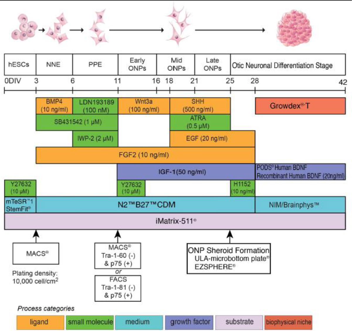
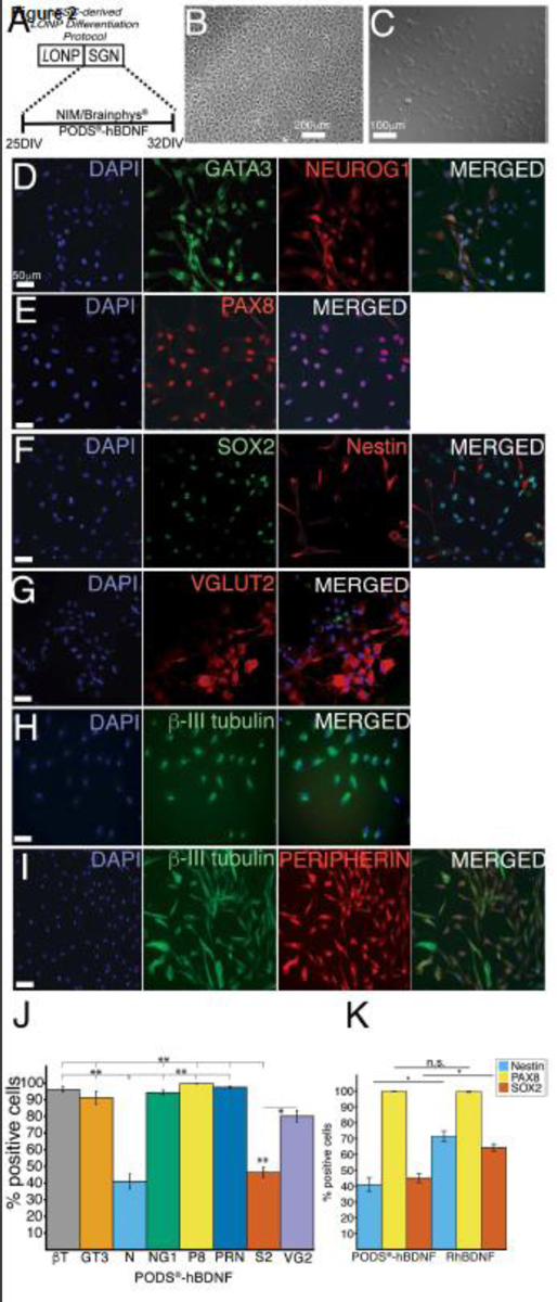
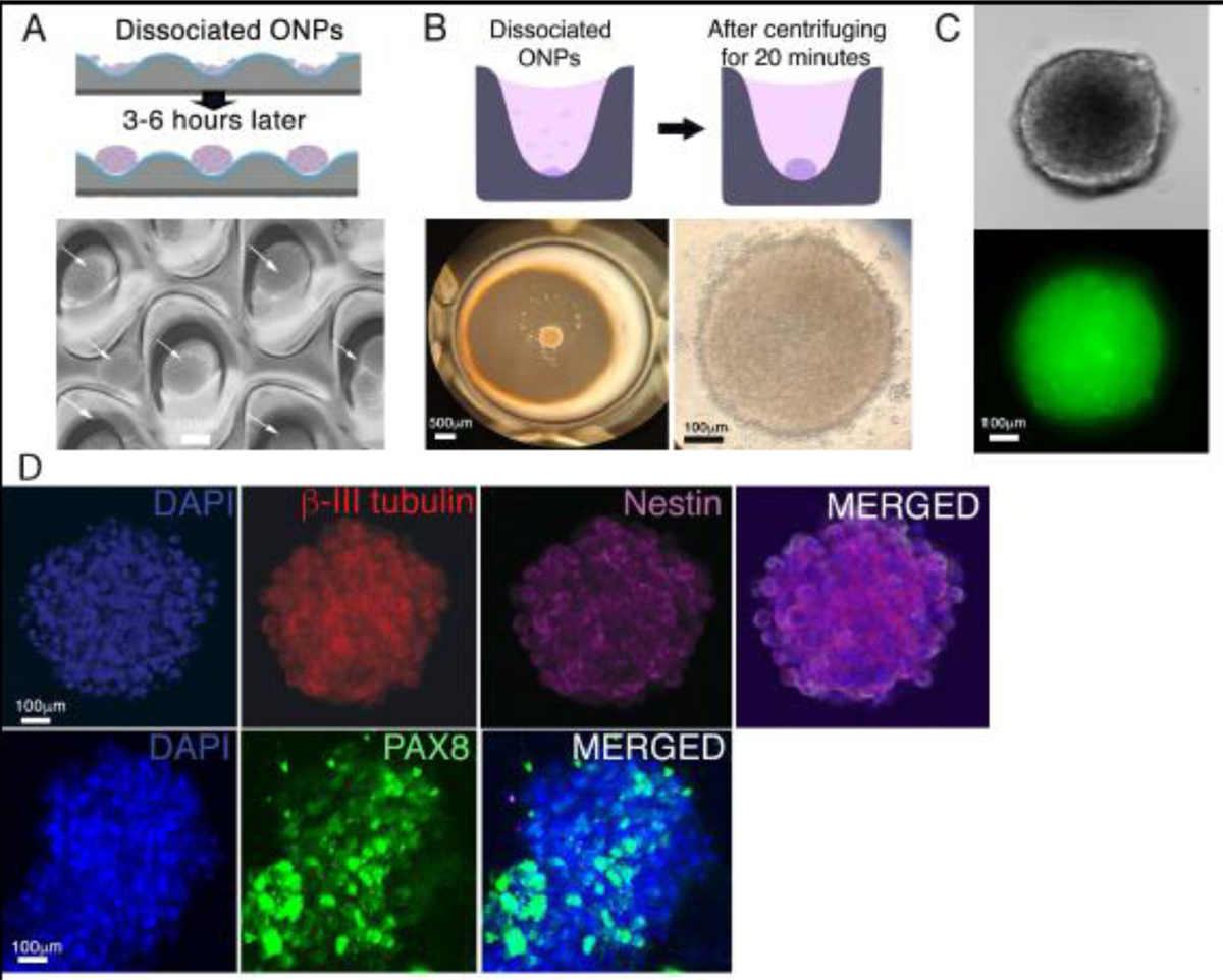
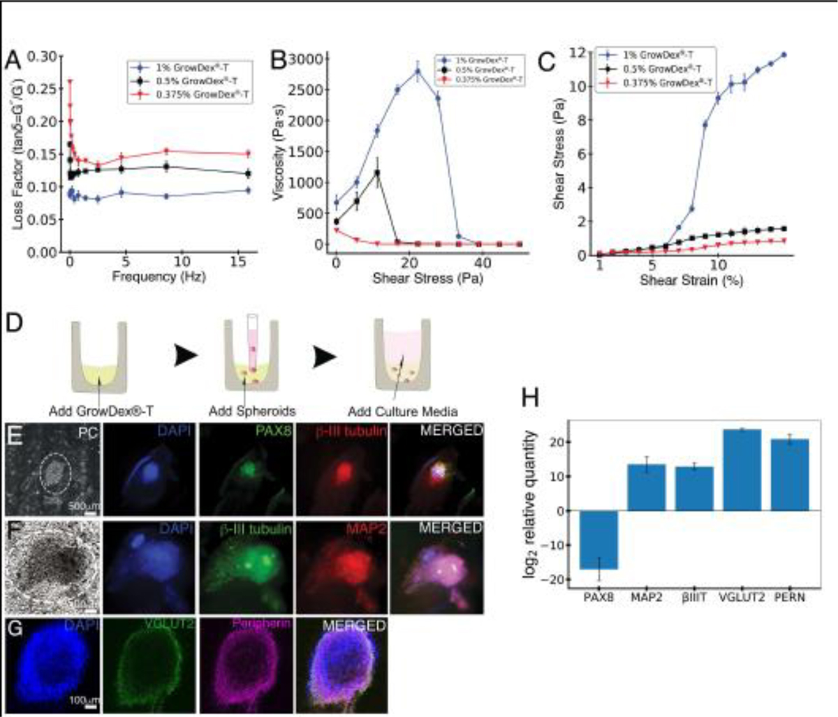
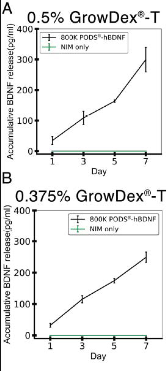
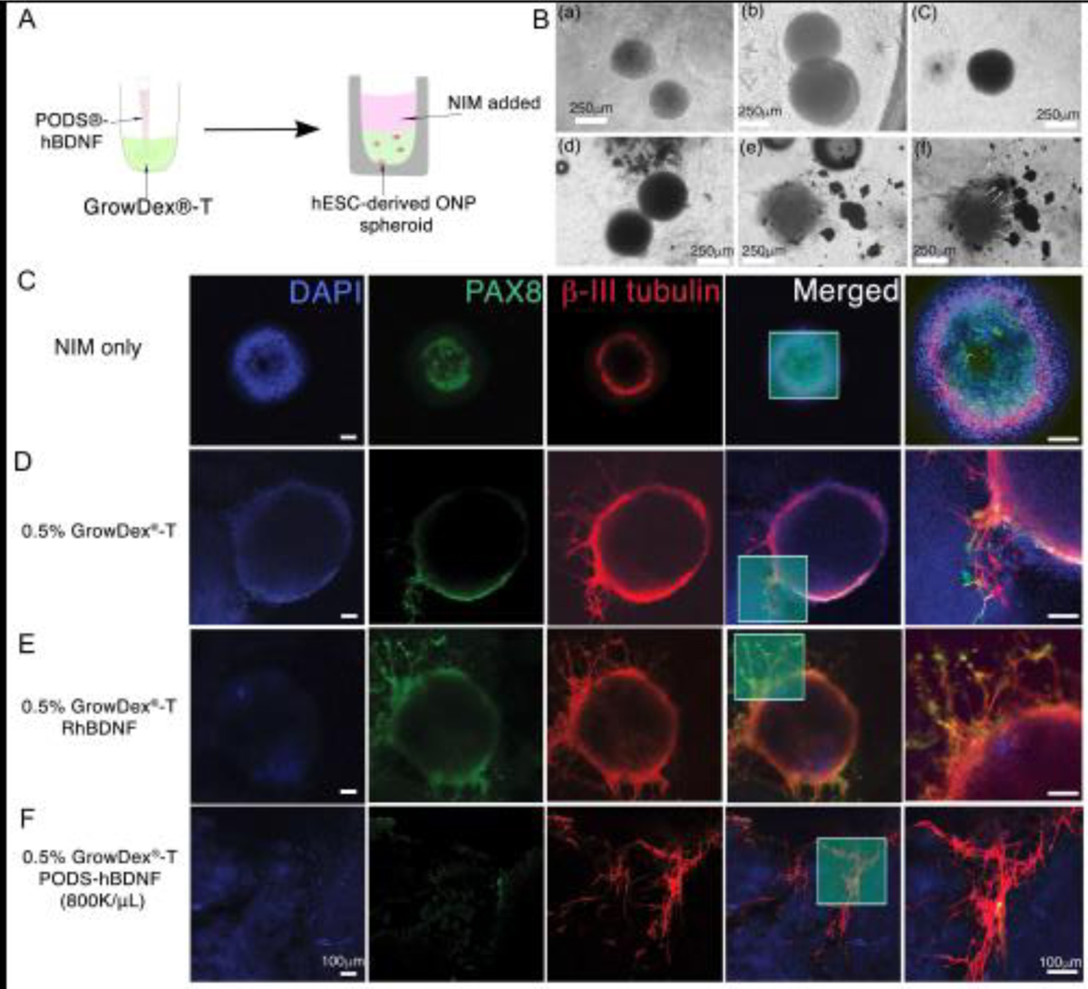
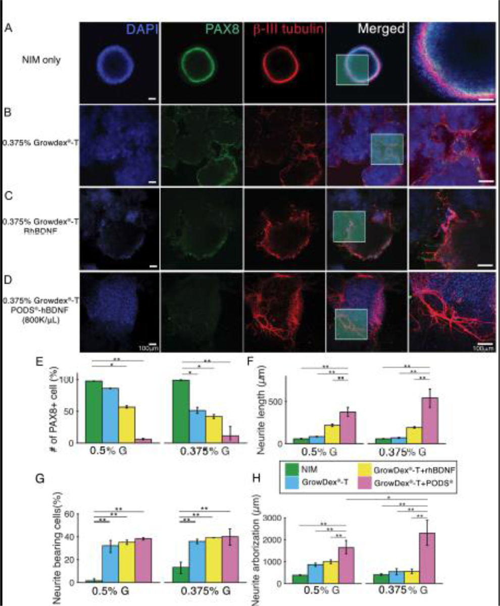
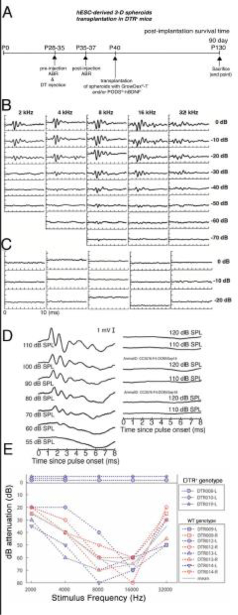
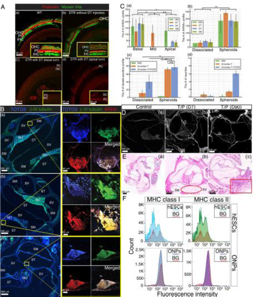
Although the application of human embryonic stem cells (hESCs) in stem cell-replacement therapy remains promising, its potential is hindered by a low cell survival rate in post-transplantation within the inner ear. Here, we aim to enhance the in vitro and in vivo survival rate and neuronal differentiation of otic neuronal progenitors (ONPs) by generating an artificial stem cell niche consisting of three-dimensional (3D) hESC-derived ONP spheroids with a nanofibrillar cellulose hydrogel and a sustained-release brain-derivative neurotrophic factor delivery system. Our results demonstrated that the transplanted hESC-derived ONP spheroids survived and neuronally differentiated into otic neuronal lineages in vitro and in vivo and also extended neurites toward the bony wall of the cochlea 90 days after the transplantation without the use of immunosuppressant medication. Our data in vitro and in vivo presented here provide sufficient evidence that we have established a robust, reproducible protocol for in vivo transplantation of hESC-derived ONPs to the inner ear. Using our protocol to create an artificial stem cell niche in the inner ear, it is now possible to work on integrating transplanted hESC-derived ONPs further and also to work toward achieving functional auditory neurons generated from hESCs. Our findings suggest that the provision of an artificial stem cell niche can be a future approach to stem cell-replacement therapy for inner-ear regeneration. STATEMENT OF SIGNIFICANCE: Inner ear regeneration utilizing human embryonic stem cell-derived otic neuronal progenitors (hESC-derived ONPs) has remarkable potential for treating sensorineural hearing loss. However, the local environment of the inner ear requires a suitable stem cell niche to allow hESC-derived ONP engraftment as well as neuronal differentiation. To overcome this obstacle, we utilized three-dimensional spheroid formation (direct contact), nanofibrillar cellulose hydrogel (extracellular matrix), and a neurotrophic factor delivery system to artificially create a stem cell niche in vitro and in vivo. Our in vitro and in vivo data presented here provide sufficient evidence that we have established a robust, reproducible protocol for in vivo transplantation of hESC-derived ONPs to the inner ear.

Keywords: Human embryonic stem cells; Human pluripotent stem cells; Hydrogel; Spiral ganglion neurons; Stem cell niche; Stem cell–replacement therapy; The inner ear.


Conflict of interest statement

Declaration of Competing Interest The authors declare that they have no known competing financial interests or personal relationships that could have appeared to influence the work reported in this paper.

Figures

Figure 1.
Figure 1.
Illustrative summary of a stepwise treatment protocol for deriving an otic neuronal lineage from undifferentiated H1, H7, and H9 human embryonic stem cells lines. Developmental stages are shown at the top, above a timeline, and key treatments are shown below, using color-coded bars. Abbreviations: D: days; NNE: nonneuronal ectoderm; PPE: preplacodal ectoderm; ONP: otic neuronal progenitor; BMP4: bone morphogenetic protein 4; SHH: Sonic hedgehog; ATRA: all-trans retinoic acid; EGF: epidermal growth factor; BDNF: brain-derived neurotrophic factor; NT3: neurotrophin 3; IGF-1: insulin-like growth factor 1; FGF2: fibroblast growth factor 2; CDM: chemically-defined medium; NIM: neural induction medium; MACS: magnetic-activated cell sorting; FACS: fluorescence-activated cell sorting; p75: low-affinity neurotrophin receptor (p75NTR). This protocol is adapted from Matsuoka et. al., 2017 and 2018.
Figure 2.
Figure 2.
Assessment of induction of an otic neuronal lineage from late-stage ONPs. (A): A stepwise treatment for an otic neuronal lineage induction. On day 25, PODS®-hBDNF treatment was started in NIM for 7 days. D: days. (B): Phase-contrast photomicrographs of hESC-derived late-stage ONPs. (C): Phase-contrast photomicrographs of hESC-derived ONPs treated with PODS®-hBDNF for seven days. (D–I): Immunocytochemistry of hESC-derived late-stage ONPs treated with PODS®-hBDNF in NIM/Brainphys™ shows expression of various otic neuronal markers: GATA3, NEUROG1, PAX8, SOX2, nestin, VGLUT2, β-III tubulin, and peripherin. (J): Quantification of the otic neuronal markers for % positivity (n = 3) on hESC-derived late-stage ONPs treated with PODS®-hBDNF for seven days. BT: β-III tubulin; GT3: GATA3; N: nestin; NG1: neurogenin 1; P8: PAX8; PRN: peripherin; S2: SOX2; and VG2: VGLUT2. (K): Quantification of % positive staining for nestin, PAX8, and SOX2 on PODS®-hBDNF and RhBDNF treated cells. *p <0.05, ** p < 0.01 by one-way ANOVA with Tukey’s post-hoc test. n.s.: not statistically significant.
Figure 3.
Figure 3.
Generation of hESC-derived ONP spheroids. (A): A schematic diagram of forming hESC-derived late-stage ONP spheroids using an EZSHPERE® plate (upper row) and a phase-contrast photomicrograph of individual spheroids (see white arrow) within the plate (bottom row). (B): A schematic diagram of forming hESC-derived ONP spheroids using a 96-well Clear Round Bottom Ultra-Low Attachment Microplate® (upper row) and a low-power (left bottom row) and high-power (right bottom row) phase-contrast photomicrograph of a spheroid within the plate. (C): A phase-contrast (upper row) and an epifluorescence (bottom row) photomicrographic image of a hESC-derived ONP spheroid stained with NeuroFluor™ NeuO. (D): Immunocytochemistry on a hESC-derived ONP spheroid that was cultured for seven days with 800,000 of PODS®-hBDNF. The spheroid was stained for β-III tubulin, nestin (upper row), and PAX8 (lower row).
Figure 4.
Figure 4.
Rheological characterization of GrowDex®-T and hESC-derived ONP spheroids cultured with GrowDex®-T. (A): Frequency sweep (viscoelasticity) of three different concentrations (1%, 0.5%, and 0.375%) of GrowDex®-T (n = 3). (B): Influence of shear stress on the viscosity of three different concentrations (1%, 0.5%, and 0.375%) of GrowDex®-T (n = 3). (C): Strain sweep (stress-strain curve) of three different concentrations (1%, 0.5% and 0.375%) of GrowDex®-T. Shear modulus of 1%, 0.5% and 0.375% GrowDex®-T is plotted as a function of shear strain (n = 3). (D): Schematic figure of hESC-derived ONP spheroids cultured in GrowDex®-T. (E–G): Neuronal marker expressions of PAX8, β-III tubulin, MAP2, VGLUT2, peripherin and phase-contrast of hESC-derived ONP spheroids cultured with 0.375% GrowDex®-T. (H): Quantitative real-time PCR (qRT-PCR) on hESC-derived ONP spheroids cultured with 0.375% GrowDex®-T and 800,000 of PODS®-hBDNF relative to hESC-derived ONP spheroids cultured in Brainphys™ with 20 ng/mL of recombinant BDNF.
Figure 5.
Figure 5.
Accumulative release kinetics of BDNF from PODS®-hBDNF and from a hESC-derived ONP spheroid cultured in NIM. (A): BDNF release profile of 800,000 PODS®-hBDNF in 0.5% GrowDex®-T over seven days. (B): BDNF release profile of 800,000 PODS®-hBDNF in 0.375% GrowDex®-T over seven days. In both (A) and (B), accumulative release kinetic of BNNF from a hESC-derived ONP spheroid cultured in NIM is also plotted in a green line as a control.
Figure 6.
Figure 6.
Immunocytochemistry of hESC-derived ONP spheroids that were cultured with 0.5% GrowDex®-T in vitro. (A): Schematic diagram of PODS®-hBDNF and GrowDex®-T mixture with hESC-derived ONPs spheroids in vitro. (B): A phase-contrast photomicrograph of hESC-derived ONP spheroids cultured with GrowDex®-T (gray background) and/or PODS®-hBDNF (shown in black). Human ESC-derived ONP spheroids that were cultured with 1% GrowDex®-T for 24 hours (a), 48 hours (b), and 72 hours (c), respectively. Human ESC-derived ONP spheroids that were cultured with 1% GrowDex®-T and 800,000/μL of PODS®-hBDNF for 24 hours (d), 48 hours (e), and 72 hours (f), respectively. (C): Immunocytochemistry of hESC-derived ONP spheroids cultured in NIM for 7 days. (D): Immunocytochemistry of hESC-derived ONP spheroids cultured in 0.5% GrowDex®-T for seven days. (E): Immunocytochemistry of hESC-derived ONP spheroids cultured in 0.5% GrowDex®-T with 20 ng/mL of RhBDNF for seven days. (F): Immunocytochemistry of hESC-derived ONP spheroids cultured in 0.5% GrowDex®-T with 800,000 of PODS®-hBDNF for 7 days.
Figure 7.
Figure 7.
Immunocytochemistry of hESC-derived ONP spheroids cultured with 0.375% GrowDex®-T in vitro. (A): Immunocytochemistry of hESCs-derived ONPs spheroids cultured in NIM for seven days. (B): Immunocytochemistry of hESC-derived ONP spheroids cultured in 0.375% GrowDex®-T for seven days. (C): Immunocytochemistry of hESC-derived ONP spheroids cultured in 0.375% GrowDex®-T with 20 ng/mL of RhBDNF for seven days. (D): Immunocytochemistry of hESC-derived ONP spheroids cultured in 0.375% GrowDex®-T with 800,000 of PODS®-hBDNF for seven days. (E): Quantification of positive staining for PAX8 based on immunocytochemistry in hESC-derived ONP spheroids. (F): Quantification of neurite length arising from hESC-derived ONP spheroids (n = 3). (G): Quantification of neurite bearing analysis. (H): Quantification of neurite arborization analysis. *p < 0.05, ** p < 0.01, *** p < 0.001 by one-way ANOVA with Tukey’s post-hoc test.
Figure 8.
Figure 8.
In vivo hESC-derived ONP spheroid transplantation in the DTR mouse cochlea. (A): A schematic diagram of experimental design for in vivo hESC-derived ONP spheroid transplantation. (B): Tone-burst-evoked ABR assessment of a WT mouse. (C): Tone-burst-evoked ABR assessment of a DTR mouse. (D): Click-evoked ABR assessment of WT mouse (left column) and DTR mouse (right column). (E): ABR audiograms for 3 DTR and 8 WT mouse ears, with mean values for the latter shown with the gray line. Levels are expressed in terms of attenuation, with sound pressure levels at 0 dB ranging from 105 to 120 dB SPL. Thresholds were based upon visual detection of a response above the noise floor. No ABRs were detected for the three DTR mice (data plotted above the 0 dB line).
Figure 9.
Figure 9.. (A):
Immunohistochemistry of the DTR mouse cochlea. Confocal fluorescence imaged of phalloidin (red) and myosin VIIa (green) in the whole mounted auditory epithelium. (a) A wild type cochlea (control) shows a single row of IHCs and three rows of OHCs in the basal turn. (b) A DTR mouse cochlea without DT injection also shows a single row of IHCs and three rows of OHCs in the basal turn as well. (c and d) A DTR mouse cochlea with DT injection at P25 demonstrates no IHCs or OHCs in the basal turn (c) and the apical turn (d). OHC: outer hair cells; IHC: inner hair cells; PC: Pillar cells, DC: Deiters cells, and ISC: inner supporting cells. B): Immunohistochemistry of transplanted hESC-derived ONP spheroids in the DTR mouse cochlea. Left column: a corresponding low-power magnification microphotograph of the DTR mouse cochlea (10X). Each yellow circle indicates the anatomical location of a transplanted hESC-derived ONP spheroid. Right column: high-power magnification microphotographs of transplanted hESC-derived ONP spheroids stained with TOTO3 (nuclear counterstaining), β-III tubulin, and AHNA (40X). (a) Human ESC-derived ONP spheroids transplanted with 1% GrowDex®-T. (b) Human ESC-derived ONP spheroids transplanted with 1% GrowDex®-T and 800,000 of PODS®-hBDNF. Small white arrow: neurites. (c) Another hESC-derived ONP spheroid transplanted with 1% GrowDex®-T and 800,000 of PODS®-hBDNF. SM: scala media, SV: scala vestibuli, ST: scala tympani, MT: the middle turn of the cochlea, and MO: modiolus. (C): (a) Quantification of the number of ANHA positive cells (profile) in three different turns of the DTR mouse cochleae. (b) Quantification of the number of the ANHA positive cells (profile) in dissociated ONPs transplantation vs. ONP spheroids transplantation in four anatomical subdivisions of the cochlea. (c) Quantification of the number of the triple positive cells (profile) in dissociated ONPs transplantation vs. ONP spheroid transplantation with three different substrates. (d): Quantification of the number of neurites in dissociated ONPs transplantation vs. ONP spheroid transplantation. NIM: neuronal induction media, G: GrowDex®-T, G+P: GrowDex®-T and PODS®-hBDNF. *p < 0.05, ** p < 0.01, N.S.: not significant by one-way ANOVA with Tukey’s post-hoc test. Note that all of the images stained for TOTO3 iodide and AHNA have been pseudo-colored. (D): Calcofluor White staining indicates the presence of NFC at both seven days and ninety days post-transplantation. (E): Tissue response in H&E histology. (a) Control WT cochlea (no surgery). (b) DTR mouse cochlea transplanted with hESC-derived ONP spheroids with GrowDex®-T and PODS®-hBDNF. The DTR mouse was euthanized seven days after the transplant surgery (D10). (c) DTR mouse cochlea transplanted with hESC-derived ONP spheroids with GrowDex®-T and PODS®-hBDNF. The DTR mouse was euthanized 90 days after the transplant surgery (D90). (F): Expression of MHC class I and MHC class II proteins assessed by flow cytometry in undifferentiated hESCs (H9) and hESC-derived ONP spheroids (ONPs). Red lines represent background staining with the conjugated antibody (MHC I and MHC II) alone. Three independent experiments were performed and the results were averaged for each analysis.

Similar articles

Cited by

References

    1. Lin FR, Niparko JK, Ferrucci L, Hearing loss prevalence in the United States, Arch Otolaryngol Head Neck Surg. 171 (2014) 2011–2012. doi:10.1002/gps.2627.6. - DOI - PMC - PubMed
    1. Goman AM, Lin FR, Prevalence of hearing loss by severity in the United States, Am. J. Public Health. 106 (2016) 1820–1822. doi:10.2105/AJPH.2016.303299. - DOI - PMC - PubMed
    1. Hinojosa R, Marion M, Histopathology of profound sensorineural deafness, Ann. N. Y. Acad. Sci 405 (1983) 459–484. doi:10.1111/j.1749-6632.1983.tb31662.x. - DOI - PubMed
    1. Glueckert R, Bitsche M, Miller JM, Zhu Y, Prieskorn DM, Altschuler RA, Schrott-Fischer A, Deafferentiation-associated changes in afferent and efferent processes in the guinea pig cochlea and afferent regeneration with chronic intrascalar brain-derived neurotrophic factor and acidic fibroblast growth factor, J. Comp. Neurol 507 (2008) 1602–1621. doi:10.1002/cne. - DOI - PubMed
    1. Spoendlin H, Factors inducing retrograde degeneration of the cochlear nerve, Ann. Otol. Rhinol. Laryngol 93 (1984) 76–82. doi:10.3109/00016487509124683. - DOI - PubMed

Publication types