. 2020 Mar 24;117(12):6726-6732.
doi: 10.1073/pnas.1913433117. Epub 2020 Mar 10.

Splice variant of growth hormone-releasing hormone receptor drives esophageal squamous cell carcinoma conferring a therapeutic target

Affiliations

Splice variant of growth hormone-releasing hormone receptor drives esophageal squamous cell carcinoma conferring a therapeutic target

Xiao Xiong et al. Proc Natl Acad Sci U S A. .

Abstract

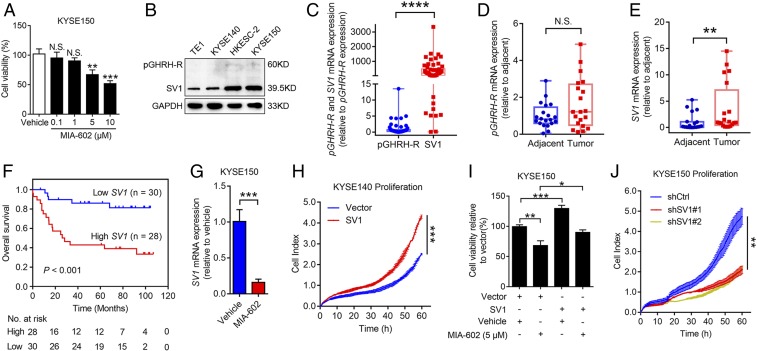
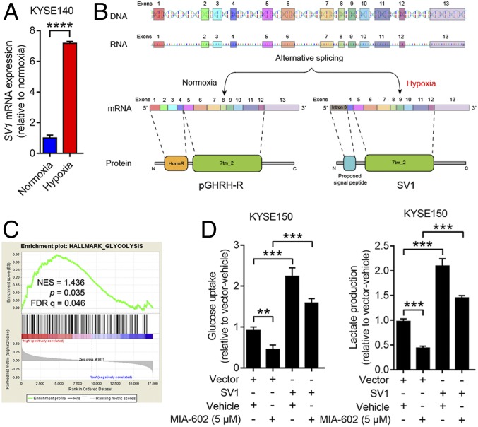
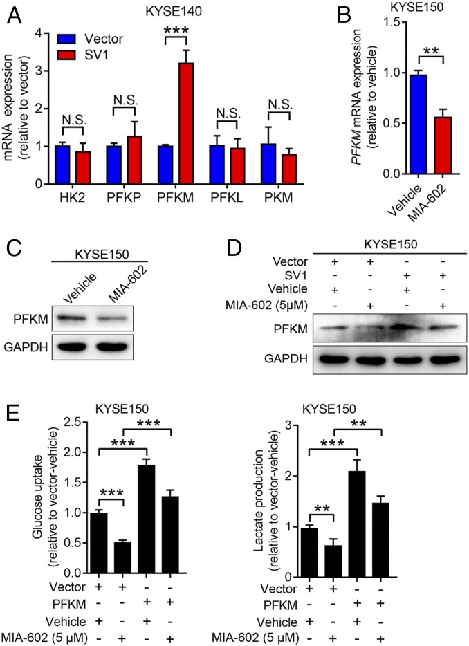
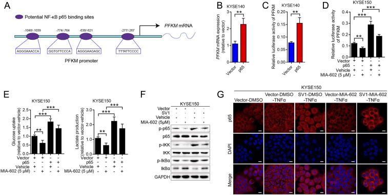
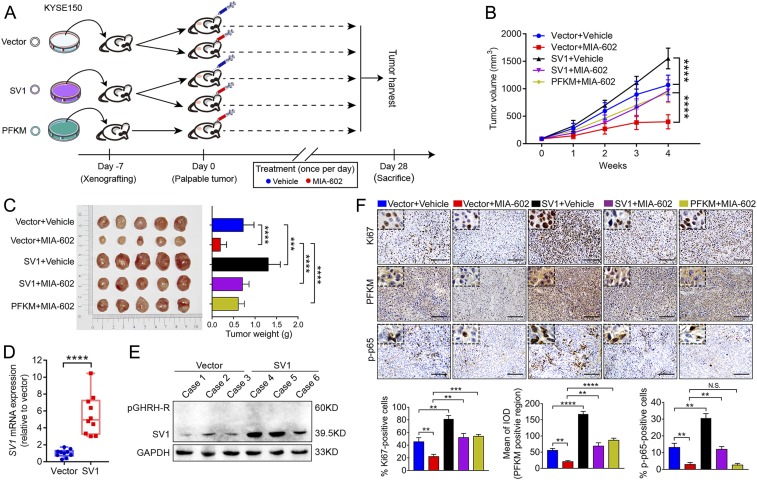
The extrahypothalamic growth hormone-releasing hormone (GHRH) and its cognate receptors (GHRH-Rs) and splice variants are expressed in a variety of cancers. It has been shown that the pituitary type of GHRH-R (pGHRH-R) mediates the inhibition of tumor growth induced by GHRH-R antagonists. However, GHRH-R antagonists can also suppress some cancers that do not express pGHRH-R, yet the underlying mechanisms have not been determined. Here, using human esophageal squamous cell carcinoma (ESCC) as a model, we were able to reveal that SV1, a known splice variant of GHRH-R, is responsible for the inhibition induced by GHRH-R antagonist MIA-602. We demonstrated that GHRH-R splice variant 1 (SV1) is a hypoxia-driven promoter of tumor progression. Hypoxia-elevated SV1 activates a key glycolytic enzyme, muscle-type phosphofructokinase (PFKM), through the nuclear factor kappa B (NF-κB) pathway, which enhances glycolytic metabolism and promotes progression of ESCC. The malignant actions induced by the SV1-NF-κB-PFKM pathway could be reversed by MIA-602. Altogether, our studies demonstrate a mechanism by which GHRH-R antagonists target SV1. Our findings suggest that SV1 is a hypoxia-induced oncogenic promoter which can be an alternative target of GHRH-R antagonists.

Keywords: GHRH-R antagonist; PFKM; glycolysis; hypoxia; splicing isoform of GHRH-R.


Conflict of interest statement

Competing interest statement: The Sponsor declares a conflict of interest. A.V.S. is a coinventor on the patent for growth hormone-releasing hormone analogs, assigned to the University of Miami, Miami, FL, and the Veterans Affairs Medical Center, Miami, FL. The other authors declare no conflict of interest.

Figures

Fig. 1.
Fig. 1.
MIA-602 inhibits ESCC cell progression through SV1. (A) Viability of KYSE150 cells treated with MIA-602 (0.1, 1, 5, or 10 μM) or vehicle solution for 48 h measured by cell-counting kit 8 (CCK-8) assay. (B) Protein levels of pGHRH-R and SV1 in four ESCC cells. GAPDH was used as an internal control. (C) RT-qPCR analyses of pGHRH-R and SV1 in 58 human ESCC specimens. (D and E) RT-qPCR analyses of pGHRH-R (D) and SV1 (E) in 20 human ESCC tissues and paired adjacent noncancerous tissues. (F) Kaplan–Meier analysis showing that OS was significantly better in patients with low expression of SV1 than in those with high expression. (G) KYSE150 cells treated with MIA-602 or vehicle; levels of SV1 were determined by RT-qPCR. (H) Proliferation of SV1-overexpressing KYSE140 cells monitored by the xCELLigence RTCA dual-plate (DP) system (ACEA Biosciences). Quantitative analysis of the cell index at 60 h is shown. (I) Viability of SV1-overexpressing KYSE150 cells treated with MIA-602 or vehicle measured by CCK-8 assay. (J) Proliferation of SV1-knockdown KYSE150 cells monitored by the xCELLigence RTCA DP system. Quantitative analysis of the cell index at 60 h is shown. Error bars indicate SEM. N.S., not significant; *P < 0.05, **P < 0.01, ***P < 0.001, ****P < 0.0001 by one-way ANOVA with post hoc intergroup comparisons (A, I, and J) or student’s t test (CE, G, and H); n = 3 in each group (A, G, and I).
Fig. 2.
Fig. 2.
Hypoxia-induced SV1 enhances glycolysis. (A) Expression of SV1 measured by RT-qPCR in KYSE140 cells pretreated at normoxia or hypoxia for 24 h. (B) Schematic of full-length GHRH-R and SV1 structures (DNA, mRNA, and protein). (C) SV1 expression positively correlates with the glycolysis pathway according to a GSEA plot (GSE47404, n = 71). (D) Glucose uptake and lactate production measured in SV1-overexpressing cells treated with MIA-602 or vehicle. Error bars indicate SEM. **P < 0.01, ***P < 0.001, ****P < 0.0001 by student’s t test (A) or one-way ANOVA with post hoc intergroup comparisons (D); n = 3 in each group (A and D).
Fig. 3.
Fig. 3.
SV1 regulates glycolysis through PFKM. (A) mRNA levels of five key glycolytic enzymes (HK2, three isoforms of PFK, and PKM) in SV1-overexpressing KYSE140 cells determined by RT-qPCR. (B and C) Expression of PFKM in KYSE150 cells treated with MIA-602 for 48 h and analyzed by RT-qPCR (B) and immunoblotting (C). GAPDH was used as an internal control. (D) Expression of PFKM in SV1-overexpressing cells treated with MIA-602 or vehicle and analyzed by immunoblotting. GAPDH was used as an internal control. (E) Glucose uptake and lactate production measured in PFKM-overexpressing cells treated with MIA-602 or vehicle. Error bars indicate SEM. N.S., not significant; **P < 0.01, ***P < 0.001 by student’s t test (A and B) or one-way ANOVA with post hoc intergroup comparisons (E); n = 3 in each group (A, B, and E).
Fig. 4.
Fig. 4.
Cancer cell glycolysis regulated by SV1-PFKM is NF-κB dependent. (A) Graphic representation of the four putative NF-κB p65 binding sites in the PFKM proximal promoter. (B) mRNA level of PFKM in p65-overexpressing cells determined by RT-qPCR. (C) The PFKM luciferase reporter was transfected into KYSE140-p65 cells and control vector cells, and the relative PFKM promoter activities were measured based on the luciferase activities. (D and E) Relative PFKM promoter activities (D), glucose uptake, and lactate production (E) measured in p65-overexpressing cells treated with MIA-602 or vehicle. (F) SV1-overexpressing cells treated with MIA-602 or vehicle before being harvested for immunoblot analyses of the labeled antigens. GAPDH was used as an internal control. (G) Subcellular localization of p65 (red) in KYSE150 cells as analyzed by immunofluorescence assay. Nuclei stained with DAPI (blue). Error bars indicate SEM. **P < 0.01, ***P < 0.001 by student’s t test (B and C) or one-way ANOVA with post hoc intergroup comparisons (D and E); n = 3 in each group (BE). (Scale bars, 10 μm.)
Fig. 5.
Fig. 5.
MIA-602 suppresses SV1-mediated ESCC tumor growth in vivo. (A) Scheme indicating the timing of xenografting and longitudinal treatment. (B) Tumor growth curves of KYSE150-SV1 and KYSE150-PFKM cells treated with MIA-602 or vehicle. (C) Tumors harvested on day 28 (Left) and average weights of tumors (Right). (D and E) Expression of SV1 detected by RT-qPCR (D) and immunoblotting (E) in tumor tissues from mice bearing KYSE150-SV1 or KYSE150-Vector cells. GAPDH was used as an internal control. (F) Representative images of immunohistochemistry of Ki67, PFKM, and p-p65 in tumor sections derived from mice (Top). Plots of percentages or mean of integrated optical density (IOD) of five groups of cells expressing the indicated proteins (Bottom). Error bars indicate SEM. N.S., not significant; **P < 0.01, ***P < 0.001, ****P < 0.0001 by one-way ANOVA with post hoc intergroup comparisons; n = 10 in each group. (Scale bars, 50 μm.)

References

    1. Oltean S., Bates D. O., Hallmarks of alternative splicing in cancer. Oncogene 33, 5311–5318 (2014). - PubMed
    1. Lin Y., et al. , Evaluation of salivary exosomal chimeric GOLM1-NAA35 RNA as a potential biomarker in esophageal carcinoma. Clin. Cancer Res. 25, 3035–3045 (2019). - PubMed
    1. Zhang H., et al. , Aberrant chimeric RNA GOLM1-MAK10 encoding a secreted fusion protein as a molecular signature for human esophageal squamous cell carcinoma. Oncotarget 4, 2135–2143 (2013). - PMC - PubMed
    1. Sibley C. R., Blazquez L., Ule J., Lessons from non-canonical splicing. Nat. Rev. Genet. 17, 407–421 (2016). - PMC - PubMed
    1. Dvinge H., Kim E., Abdel-Wahab O., Bradley R. K., RNA splicing factors as oncoproteins and tumour suppressors. Nat. Rev. Cancer 16, 413–430 (2016). - PMC - PubMed

Publication types

MeSH terms

Substances

LinkOut - more resources