. 2020 Mar 10;11(1):1299.
doi: 10.1038/s41467-020-14336-7.

Decorating bacteria with self-assembled synthetic receptors

Affiliations

Decorating bacteria with self-assembled synthetic receptors

Naama Lahav-Mankovski et al. Nat Commun. .

Abstract

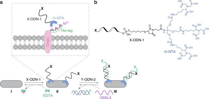
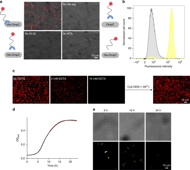
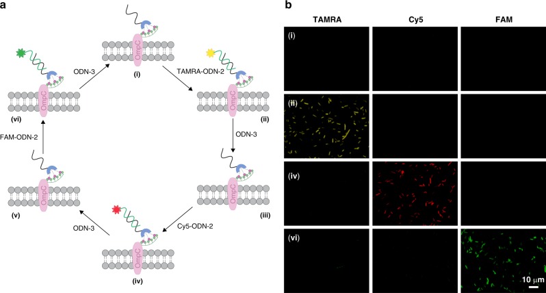
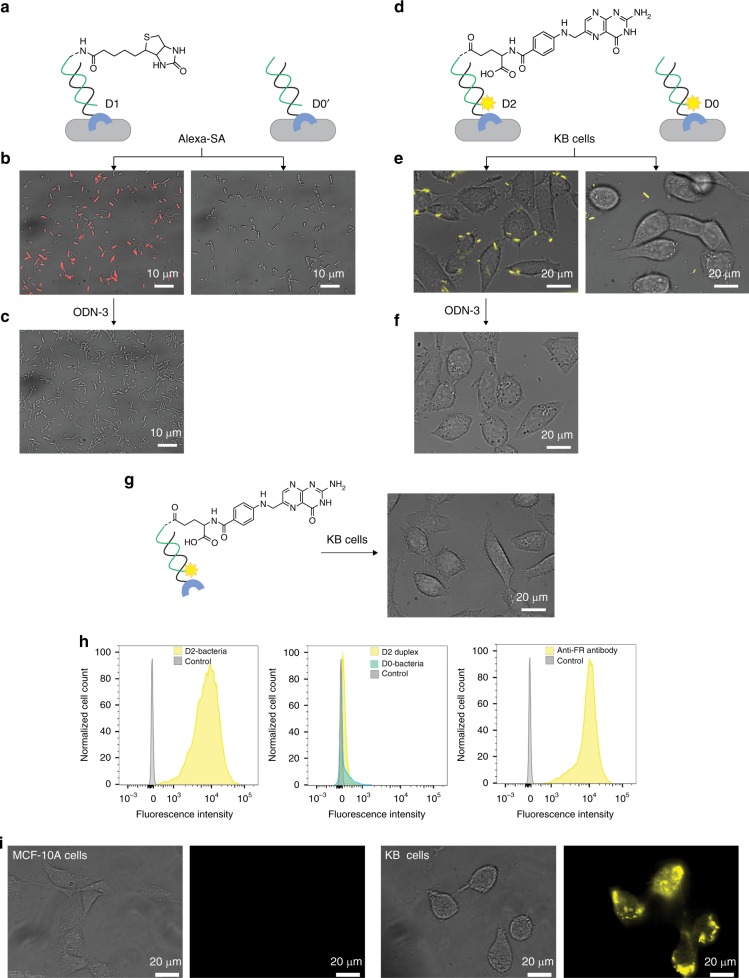
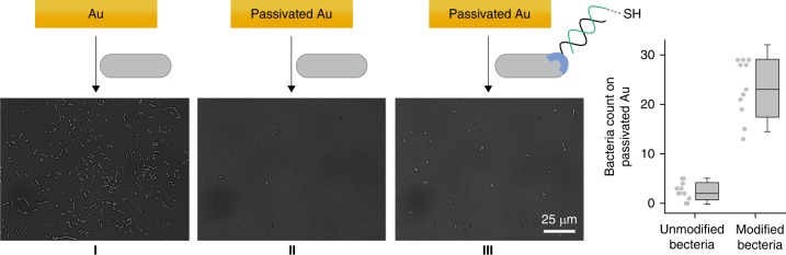
The responses of cells to their surroundings are mediated by the binding of cell surface proteins (CSPs) to extracellular signals. Such processes are regulated via dynamic changes in the structure, composition, and expression levels of CSPs. In this study, we demonstrate the possibility of decorating bacteria with artificial, self-assembled receptors that imitate the dynamic features of CSPs. We show that the local concentration of these receptors on the bacterial membrane and their structure can be reversibly controlled using suitable chemical signals, in a way that resembles changes that occur with CSP expression levels or posttranslational modifications (PTMs), respectively. We also show that these modifications can endow the bacteria with programmable properties, akin to the way CSP responses can induce cellular functions. By programming the bacteria to glow, adhere to surfaces, or interact with proteins or mammalian cells, we demonstrate the potential to tailor such biomimetic systems for specific applications.


Conflict of interest statement

The authors declare no competing interests.

Figures

Fig. 1
Fig. 1. Design principles.
a One way to decorate E. coli with artificial receptors, which are appended with a specific functionality (X), involves the binding of X-ODN-1 to a hexa-histidine tag (His-tag) fused to OmpC (I → II). This process can be reversed by subjecting the bacteria to EDTA (II → I). Another way to introduce an unnatural recognition motif (Y) to the bacterial surface is by adding to the bacteria decorated with ODN-1 a complementary strand modified with the desired functionality (Y-ODN-2, II → III). Y-ODN-2 can be selectively removed by adding a complementary strand, ODN-3 (III → II). b Structure of X-ODN-1.
Fig. 2
Fig. 2. Reversible, non-covalent modification of a bacterial membrane using ODN-based synthetic receptors.
a Merged bright-field and fluorescence images of the following: (Top left) E. coli expressing His-OmpC incubated with 500 nM of Cy5-ODN-1 and Ni (II). (Top right) Bacteria lacking His-tag incubated with 500 nM of Cy5-ODN-1 and Ni (II). (Bottom left) His-tagged bacteria incubated with 500 nM of Cy5-ODN-1 in the absence of Ni (II). (Bottom right) His-tagged bacteria incubated with 500 nM of Cy5-ODN (that lacks the NTA group) and Ni (II). b Flow cytometry analysis of His-tagged bacteria (yellow) and bacteria lacking His-tag (gray) incubated with TAMRA-ODN-1. c Images of E. coli expressing His-OmpC decorated with Cy5-ODN-1 in the presence of increasing concentrations of EDTA (0, 5, and 10 mM) (left), and following the subsequent addition of Cy5-ODN-1 in the presence of Ni (II) (right). d Growth curve of E. coli expressing His-OmpC (black) and the growth of the same bacteria decorated with TAMRA-ODN-1 (red). e Bright-field (top) and fluorescence images (bottom) of bacteria decorated with TAMRA-ODN-1 monitored at 0, 12, and 24 h. Source data are available in the Source Data file.
Fig. 3
Fig. 3. Reversible modification of synthetic receptors bound to the bacterial membrane.
a A synthetic receptor bound to the His-tagged bacteria (ODN-1) can be sequentially modified with unnatural functionalities, here, fluorescent dyes (TAMRA, Cy5, or FAM), simply by incubating the modified bacteria with a dye-modified ODN-2 that can be selectively removed using ODN-3. b Monitoring states i, ii, iv, and vi in the sequential labeling process (the scheme on the left) by simultaneously observing the emissions of TAMRA, Cy5, and FAM.
Fig. 4
Fig. 4. Programing the bacteria to interact with proteins and cancer cells.
a Schematic illustration of an experiment in which modified His-tagged bacteria were treated with Alexa 647-modified streptavidin (Alexa-SA). Left: Bacteria modified with a D1 duplex generated from ODN-1 and biotin-ODN-2. Right: Bacteria modified with a D0’ duplex that lacks biotin. b The resulting images (merged bright-field and fluorescence). c Images recorded following the incubation of the bacteria bound to Alexa-SA with ODN-3. d Schematic illustration of an experiment in which modified His-tagged bacteria were incubated with KB-cells. Left: Bacteria decorated with a D2 duplex consisting of ODN-1 and TAMRA-labeled folate-ODN-2. Right: Bacteria decorated with a D0 duplex that lacks the folate group. e The resulting images. f Images obtained after treating the bacteria that are bound to KB cells with ODN-3. g Incubating KB-cells with the D2 duplex (in the absence of bacteria) did not lead to fluorescence labeling of the KB-cell. h Representative flow cytometry histograms of KB cells before (gray) and after (yellow/cyan) treatment with: (left) D2-modified bacteria (16 × 108 cells/ml), (middle) D2 alone or D0-modified bacteria (500 nM), and (right) anti-FR antibody (0.31 μg/ml). i Bright field and fluorescent images of healthy MCF-10A cells (left) and cancerous KB cells (right) treated with the D2-modified bacteria.
Fig. 5
Fig. 5. Programing the bacteria to adhere to surfaces.
a Images of the following: (I) bare gold substrate after incubation with unmodified bacteria, (II) passivated gold substrate after incubation with unmodified bacteria, and (III) passivated gold substrate following incubation with bacteria modified with a thiol-modified duplex (ODN-1: HS-ODN-2). b Average number of bacteria per 0.0165 mm2 of passivated gold. The error bars represent the s.d. of 11 frames. Source data are available in the Source Data file.
Fig. 6
Fig. 6. Controlling bacterial cell surface luminescence.
a Schematic illustration of the way sub-populations of His-tagged bacteria bearing artificial receptors can be prepared (I), mixed (I → II), and selectively modified in the mixture (II → III) using ODN-2s appended with FAM, TAMRA, and Cy5. Complementary sequences are denoted in similar colors. b Fluorescence image of the labeled mixed population. The image corresponds to state III in the scheme. The bacteria were imaged using 488, 561, and 647 nm excitation lasers. c Percentage of each sub-population in the mixture. The error bars represent the s.d. of six frames. d Flow cytometry analysis of the mixed population. e STORM images of His-tagged bacteria decorated with the ODN-1:Cy5-ODN-2 duplex. Left: whole bacteria. Right: transverse cut viewed from the plane of the cell axis. Source data are available in the Source Data file.

Similar articles

Cited by

References

    1. Bausch-Fluck D, et al. The in silico human surfaceome. Proc. Natl Acad. Sci. USA. 2018;115:E10988–E10997. doi: 10.1073/pnas.1808790115. - DOI - PMC - PubMed
    1. Berne, C., Ducret, A., Hardy, G. G. & Brun, Y. V. Adhesins involved in attachment to abiotic surfaces by Gram-negative bacteria. Microbiol. Spectr. 3, 10.1128/microbiolspec.MB-0018-2015 (2015). - PMC - PubMed
    1. Moorthy S, Keklak J, Klein EA. Perspective: adhesion mediated signal transduction in bacterial pathogens. Pathogens. 2016;5:23. doi: 10.3390/pathogens5010023. - DOI - PMC - PubMed
    1. Pereira CS, Thompson JA, Xavier KB. AI-2-mediated signalling in bacteria. FEMS Microbiol. Rev. 2013;37:156–181. doi: 10.1111/j.1574-6976.2012.00345.x. - DOI - PubMed
    1. Verbeke, F. et al. Peptides as quorum sensing molecules: measurement techniques and obtained levels in vitro and in vivo. Front. Neurosci. 11, 183 (2017). - PMC - PubMed

Publication types