. 2020 Apr;14(1):157-161.
doi: 10.1007/s12104-020-09937-8. Epub 2020 Mar 10.

pH-dependent secondary structure propensity of the influenza A virus M2 cytoplasmic tail

Affiliations

pH-dependent secondary structure propensity of the influenza A virus M2 cytoplasmic tail

Jolyon K Claridge et al. Biomol NMR Assign. 2020 Apr.

Abstract

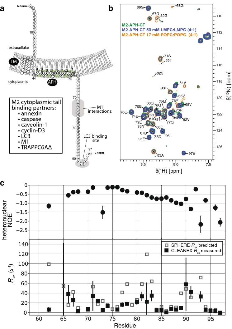
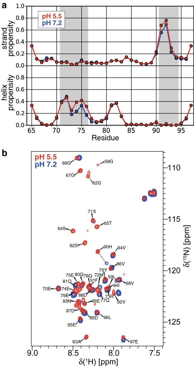
The cytoplasmic C-terminal tail of the matrix protein 2 (M2) from influenza A virus has a well conserved sequence and is involved in interactions with several host proteins as well as the influenza matrix protein 1 (M1). Whereas the transmembrane domain of M2 has been well characterised structurally and functionally, high resolution information about the distal cytoplasmic tail is lacking. Here we report the chemical shifts of the cytoplasmic tail of M2 and the chemical shift perturbations at low pH and in the presence of membrane mimetics. The cytoplasmic tail residues are mostly disordered but an extended backbone conformation is adopted by the LC3 binding motif and the putative M1 interaction site has partial helical content with a small pH-dependence. The chemical shift assignments provide a basis for further investigations into interactions of the M2 cytoplasmic tail with viral and host cell factors.

Keywords: Influenza; M2; Matrix protein 2.


Conflict of interest statement

The authors declare that they have no conflict of interest.

Figures

Fig. 1
Fig. 1
a Schematic of full-length influenza M2 with the transmembrane (TM) and membrane-attached amphipathic helix (APH) indicated. Some of the proteins that have been shown to interact with the M2 cytoplasmic tail are listed. The regions responsible for interaction with M1 and LC3 are indicated on the schematic with gray shading. The direction of membrane curvature expected along the axis parallel to the direction of the budding event is shown. b Overlay of the amide spectra for M2-APH-CT (residues 44–97) at pH 7.2 by itself (green), in the presence of LMPC:LMPG (4:1) micelles (blue), and in the presence POPC:POPG (4:1) liposomes (orange). The two crosspeaks at ~ 109 ppm and ~ 111 ppm (in 15N) and indicated with asterisks are attributed to Gly58 and Gly62 in the absence of membrane mimetics. c Top, 1H–15N heteronuclear NOEs for M2-APH-CT in the presence of POPC:POPG liposomes at pH 7.2. Bottom, amide proton water exchange rates (Rex) derived from CLEANEX experiments for M2-APH-CT in the presence of POPC:POPG liposomes at pH 7.2. Also shown are the exchange rates predicted from sequence alone using the SPHERE server (Yu-Zhu Zhang, Ph.D. Thesis, University of Pennsylvania, PA, USA.)
Fig. 2
Fig. 2
a Helix and strand propensities of M2-APH-CT at pH 7.2 and pH 5.5 as determined from chemical shifts using TALOS-N. The regions 70–72 and 91–94 are shaded to indicate the presence of secondary structure in the binding sites for M1 and LC3, respectively. b Overlay of the amide spectra of M2-APH-CT at pH 5.5 and pH 7.2 in the presence of 50 mM LMPC:LMPG (4:1). The chemical shift perturbations of H90 and V92 are emphasised with dotted lines. Both spectra were recorded at 310 K

References

    1. Beale R, Wise H, Stuart A, Ravenhill BJ, Digard P, Randow F. A LC3-interacting motif in the influenza A virus M2 protein is required to subvert autophagy and maintain virion stability. Cell Host Microbe. 2014;15(2):239–247. doi: 10.1016/j.chom.2014.01.006. - DOI - PMC - PubMed
    1. Busch DJ, Houser JR, Hayden CC, Sherman MB, Lafer EM, Stachowiak JC. Intrinsically disordered proteins drive membrane curvature. Nat Commun. 2015;6:7875. doi: 10.1038/ncomms8875. - DOI - PMC - PubMed
    1. Cady SD, Schmidt-Rohr K, Wang J, Soto CS, Degrado WF, Hong M. Structure of the amantadine binding site of influenza M2 proton channels in lipid bilayers. Nature. 2010;463(7281):689–692. doi: 10.1038/nature08722. - DOI - PMC - PubMed
    1. Chen BJ, Leser GP, Jackson D, Lamb RA. The influenza virus M2 protein cytoplasmic tail interacts with the M1 protein and influences virus assembly at the site of virus budding. J Virol. 2008;82(20):10059–10070. doi: 10.1128/JVI.01184-08. - DOI - PMC - PubMed
    1. Cho KJ, Schepens B, Seok JH, Kim S, Roose K, Lee JH, Gallardo R, Van Hamme E, Schymkowitz J, Rousseau F, Fiers W, Saelens X, Kim KH. Structure of the extracellular domain of matrix protein 2 of influenza A virus in complex with a protective monoclonal antibody. J Virol. 2015;89(7):3700–3711. doi: 10.1128/JVI.02576-14. - DOI - PMC - PubMed

Publication types

MeSH terms

Substances