Review
. 2020 Feb 25:10:61.
doi: 10.3389/fcimb.2020.00061. eCollection 2020.

HIV-1 Tat: Role in Bystander Toxicity

Affiliations
Review

HIV-1 Tat: Role in Bystander Toxicity

David Ajasin et al. Front Cell Infect Microbiol. .

Abstract

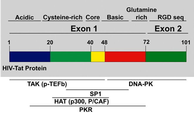
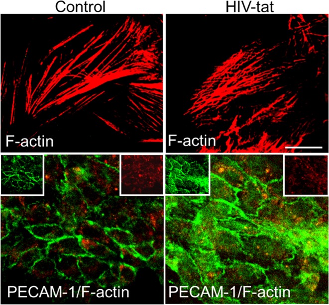
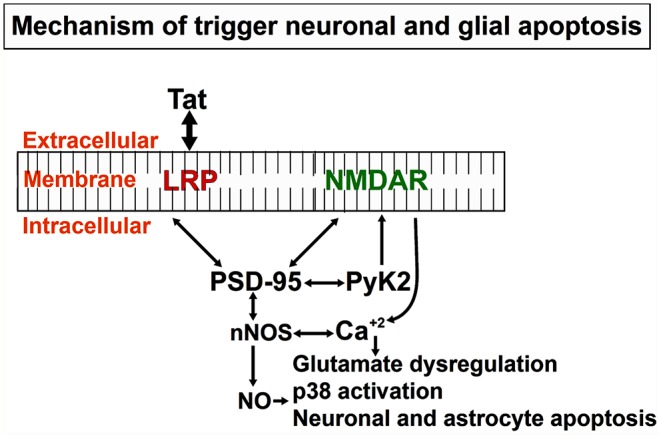
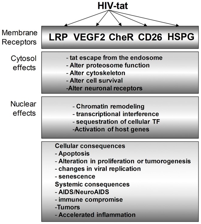
HIV Tat protein is a critical protein that plays multiple roles in HIV pathogenesis. While its role as the transactivator of HIV transcription is well-established, other non-viral replication-associated functions have been described in several HIV-comorbidities even in the current antiretroviral therapy (ART) era. HIV Tat protein is produced and released into the extracellular space from cells with active HIV replication or from latently HIV-infected cells into neighboring uninfected cells even in the absence of active HIV replication and viral production due to effective ART. Neighboring uninfected and HIV-infected cells can take up the released Tat resulting in the upregulation of inflammatory genes and activation of pathways that leads to cytotoxicity observed in several comorbidities such as HIV associated neurocognitive disorder (HAND), HIV associated cardiovascular impairment, and accelerated aging. Thus, understanding how Tat modulates host and viral response is important in designing novel therapeutic approaches to target the chronic inflammatory effects of soluble viral proteins in HIV infection.

Keywords: HIV-1; HIV-1 latency; Tat; cardiomyopathy; connexin; neuro-HIV.


Figures

Figure 1
Figure 1
HIV Tat domain structures: tat protein is encoded from exons. Exon 1 encodes for multiple domains like the cysteine-rich, core, the basic domain, and the glutamine-rich domain. Exon 2 encodes for the Arginine rich domain. Most of the p-TEFb complex interactions, as well as other interactions necessary for transcription of HIV provirus, are mediated by domains in Exon 1.
Figure 2
Figure 2
Tat alters actin cytoskeleton and PECAM expression. Cells were treated as empty media and HIV-1 Tat containing media. In control cells, actin cytoskeleton (Cy3-red) remains intact, and PECAM expression (green) is as expected mainly localized to the plasma membrane. In Tat treated samples, actin cytoskeleton is disrupted, and PECAM expression and distribution changes to be in the cytoplasm and at the plasma membrane.
Figure 3
Figure 3
Mechanistic depiction of Tat induced neuronal and astrocytic apoptosis: Tat protein taken up by bystander cells induces the formation of nNOS that leads to the formation of Nitric Oxide (NO), significant release of calcium from intracellular calcium stores that triggers other downstream mediators of apoptosis in both neurons and astrocytes. Apoptosis of both cell types (neurons and astrocytes), as well as others like axonal/dendritic shrinkage, causes a neurocognitive decline in HIV-infected individuals.
Figure 4
Figure 4
Multiple intracellular mediators of the Tat effect on bystander cells. Tat protein modulates HIV-infected cells as we as uninfected bystander cells. In the cytosol, the effect of Tat ranges from altering the proteasomal degradation pathway to favor viral and host proteins that support viral replication, cell survival, and other identified phenotypes. Similarly, Tat mediates multiple changes in the nucleus to effect transcriptional changes that can vary cellular effects such as apoptosis, inflammation, and many more.

References

    1. Abraham S., Sawaya B. E., Safak M., Batuman O., Khalili K., Amini S. (2003). Regulation of MCP-1 gene transcription by Smads and HIV-1 Tat in human glial cells. Virology 309, 196–202. 10.1016/S0042-6822(03)00112-0 - DOI - PubMed
    1. Abulwerdi F. A., Le Grice S. F. J. (2017). Recent advances in targeting the HIV-1 Tat/TAR complex. Curr. Pharm. Des. 23, 4112–4121. 10.2174/1381612823666170616081736 - DOI - PubMed
    1. Aksenova M. V., Aksenov M. Y., Adams S. M., Mactutus C. F., Booze R. M. (2009). Neuronal survival and resistance to HIV-1 Tat toxicity in the primary culture of rat fetal neurons. Exp. Neurol. 215, 253–263. 10.1016/j.expneurol.2008.10.006 - DOI - PMC - PubMed
    1. Albini A., Benelli R., Giunciuglio D., Cai T., Mariani G., Ferrini S., et al. . (1998a). Identification of a novel domain of HIV tat involved in monocyte chemotaxis. J. Biol. Chem. 273, 15895–15900. 10.1074/jbc.273.26.15895 - DOI - PubMed
    1. Albini A., Ferrini S., Benelli R., Sforzini S., Giunciuglio D., Aluigi M. G., et al. . (1998b). HIV-1 Tat protein mimicry of chemokines. Proc. Natl. Acad. Sci. U.S.A. 95, 13153–13158. 10.1073/pnas.95.22.13153 - DOI - PMC - PubMed

Publication types

Substances