. 2020 Jan 13;6(1):553-563.
doi: 10.1021/acsbiomaterials.9b01241. Epub 2019 Nov 22.

Tailored Three-Dimensionally Printed Triply Periodic Calcium Phosphate Implants: A Preclinical Study for Craniofacial Bone Repair

Affiliations

Tailored Three-Dimensionally Printed Triply Periodic Calcium Phosphate Implants: A Preclinical Study for Craniofacial Bone Repair

Arnaud Paré et al. ACS Biomater Sci Eng. .

Erratum in

Abstract

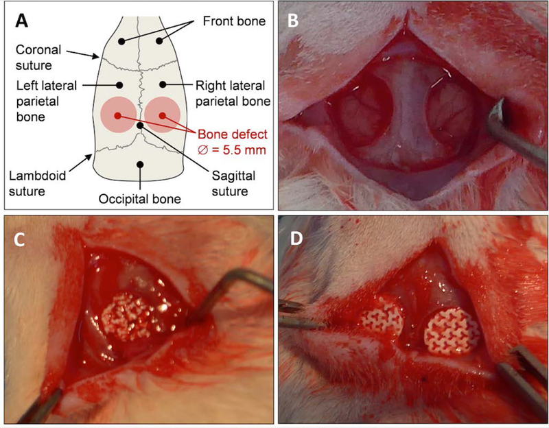
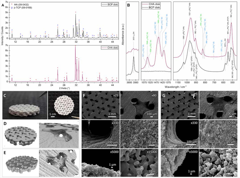
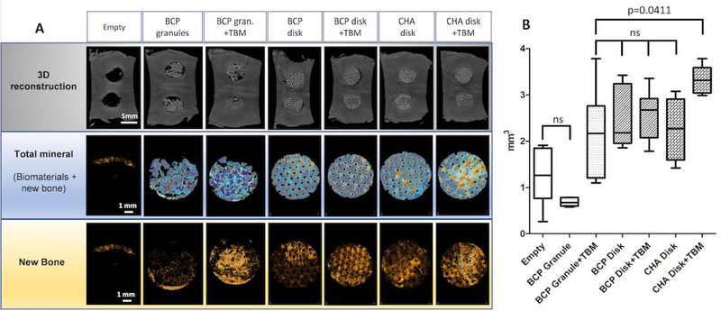
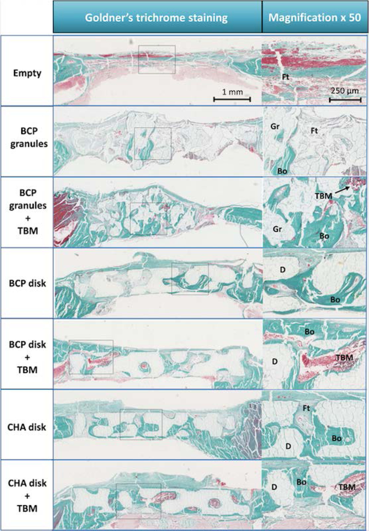
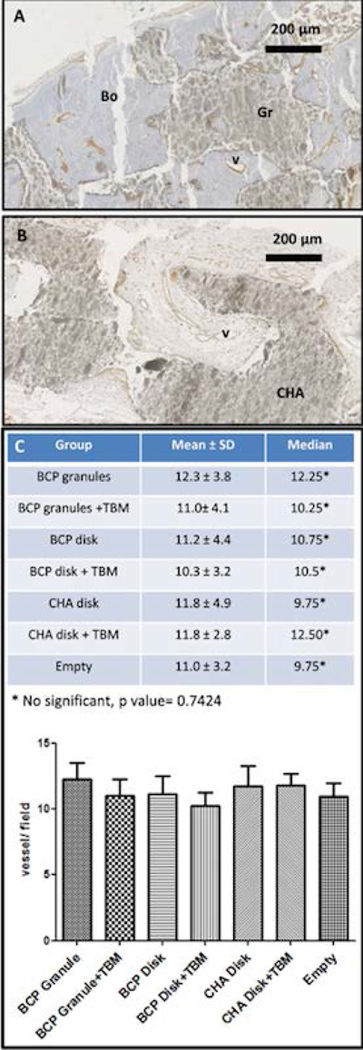
Finding alternative strategies for the regeneration of craniofacial bone defects (CSD), such as combining a synthetic ephemeral calcium phosphate (CaP) implant and/or active substances and cells, would contribute to solving this reconstructive roadblock. However, CaP's architectural features (i.e., architecture and composition) still need to be tailored, and the use of processed stem cells and synthetic active substances (e.g., recombinant human bone morphogenetic protein 2) drastically limits the clinical application of such approaches. Focusing on solutions that are directly transposable to the clinical setting, biphasic calcium phosphate (BCP) and carbonated hydroxyapatite (CHA) 3D-printed disks with a triply periodic minimal structure (TPMS) were implanted in calvarial critical-sized defects (rat model) with or without addition of total bone marrow (TBM). Bone regeneration within the defect was evaluated, and the outcomes were compared to a standard-care procedure based on BCP granules soaked with TBM (positive control). After 7 weeks, de novo bone formation was significantly greater in the CHA disks + TBM group than in the positive controls (3.33 mm3 and 2.15 mm3, respectively, P=0.04). These encouraging results indicate that both CHA and TPMS architectures are potentially advantageous in the repair of CSDs and that this one-step procedure warrants further clinical investigation.

Keywords: 3D printing; Bioceramics; Bone marrow; Bone tissue engineering; Calcium phosphates; Calvaria.


Conflict of interest statement

Conflicts of interest: None

Figures

Figure 1.
Figure 1.. Calvarial bone defect in rat model.
(A) Diagram and (B) picture showing the two critical-size calvarial defects performed on the left and right parietal bone of inbred Lewis rat (5.5 mm in diameter). Pictures showing the defects filled with (C) BCP granules and (D) macroporous disk-shaped bioceramics.
Figure 2.
Figure 2.. Design of the macroporous bioceramic disk.
3D images derived from Simpleware CAD software of (A) the macroporous disk with a 40% porosity volume, (B) cross section and top views of a 40% gyroid structure wherein a sphere 300 μm in diameter can move freely and (C) the changes in the maximum diameter of a sphere that can go through the entire gyroid structure depending on the size of the gyroid’s fundamental unit and its porosity.
Figure 3.
Figure 3.. Characteristics of macroporous bioceramics.
(A) XRD patterns and (B) IR spectra of the BCP and CHA ground scaffolds assessing the scaffold composition. “CO32− (B)” and “CO32− (A)” correspond to vibrations of carbonate ions in the positions occupied by phosphate and hydroxide ions in the HA lattice, respectively. (C) Pictures, and 3D images of the (D) BCP and (E) CHA bioceramics obtained by X-ray μ-tomography. SEM micrographs showing the macro- and micro-architecture of (F) BCP and (G) CHA bioceramics including 300 μm macropores and submicron micropores.
Figure 4.
Figure 4.. Micro-CT analysis of the critical-sized craniofacial bone defect (CSD) reconstruction.
(A) Images of the CSD reconstructions at 7 weeks showing calvarial 3D reconstruction, biomaterials + new bone as well as newly formed bone alone. CSD repair with BCP granules ± TBM systemically had biomaterial loss and calvarial holes. (B) Graph showing the quantitative analysis of bone volume (BV, mm3) in the region of interest. Empty defect (negative control) and BCP Granule groups had the lowest rate of bone formation compared to others (p<0.05). There was no statistical difference between BCP granule+TBM (positive control) and macroporous disks alone while the CHA+TBM group had significantly higher bone formation than BCP granule+TBM (p=0.0441).
Figure 5.
Figure 5.. IHC of calvarial reconstruction using Goldner’s Trichome staining.
Images showing the histological assessment at 7 weeks of CSD reconstruction with a magnification (×50) of a corresponding new bone formation area. The calvarial reconstructions using custom-made macroporous disks evenly restored the cranial vault compared to groups reconstructed by BCP granules. In groups using CaP biomaterials without TBM, more newly formed bone was observed in groups reconstructed by macroporous disks than by BCP granules. Live TBM cells were systematically observed in groups reconstructed by CaP biomaterials+TBM. A large amount of new bone was observed when biomaterials were combined with TBM. Abbreviations: new bone, Bo; BCP granule, Gr; macroporous disk, D; total bone marrow, TBM; fibrous tissues, Ft.
Figure 6:
Figure 6:. Assessment of angiogenesis in craniofacial bone defect (CSD) reconstruction.
IHC images showing vessel formation in calvarial reconstruction with (A) BCP granules or (B) CHA macroporous bioceramic using endothelial cell immunostaining with anti-CD 31 antibody. (C) Graph and table of the vessel quantification (vessel count of 10 fields per CSD, magnification ×40) showing no significant difference between experimental groups. Abbreviations: Bo, new bone; v, vessel; Gr, granules; CHA, CHA macroporous bioceramic.

References

    1. Sen MK; Miclau T Autologous Iliac Crest Bone Graft: Should It Still Be the Gold Standard for Treating Nonunions? Injury 2007, 38 (1), S75–S80. 10.1016/j.injury.2007.02.012. - DOI - PubMed
    1. Grant GA; Jolley M; Ellenbogen RG; Roberts TS; Gruss JR; Loeser JD Failure of Autologous Bone-Assisted Cranioplasty Following Decompressive Craniectomy in Children and Adolescents. J. Neurosurg. 2004, 100 (2 Suppl Pediatrics), 163–168. 10.3171/ped.2004.100.2.0163. - DOI - PubMed
    1. Honeybul S; Morrison DA; Ho KM; Lind CRP; Geelhoed E A Randomized Controlled Trial Comparing Autologous Cranioplasty with Custom-Made Titanium Cranioplasty. J. Neurosurg. 2017, 126 (1), 81–90. https://doi.org/10.317½015.12.JNS152004. - PubMed
    1. Dünisch P; Walter J; Sakr Y; Kalff R; Waschke A; Ewald C Risk Factors of Aseptic Bone Resorption: A Study after Autologous Bone Flap Reinsertion Due to Decompressive Craniotomy. J. Neurosurg. 2013, 118 (5), 1141–1147. https://doi.org/10.317½013.1.JNS12860. - PubMed
    1. Daculsi G; Baroth S; LeGeros R 20 Years of Biphasic Calcium Phosphate Bioceramics Development and Applications In Ceramic Engineering and Science Proceedings; Narayan R, Colombo P, Singh D, Salem J, Eds.; John Wiley & Sons, Inc.: Hoboken, NJ, USA, 2009; pp 45–58. 10.1002/9780470584354.ch5. - DOI

Publication types

Substances

LinkOut - more resources