Review
. 2019 May 25:1:100008.
doi: 10.1016/j.mtbio.2019.100008. eCollection 2019 Jan.

Bioinks and bioprinting technologies to make heterogeneous and biomimetic tissue constructs

Affiliations
Review

Bioinks and bioprinting technologies to make heterogeneous and biomimetic tissue constructs

N Ashammakhi et al. Mater Today Bio. .

Abstract

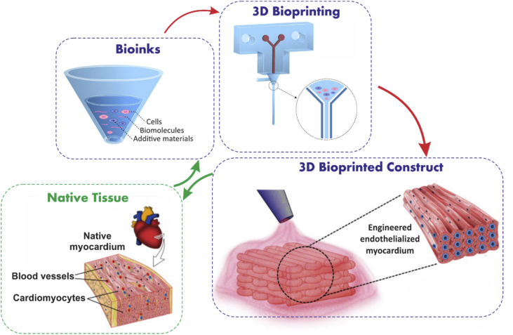
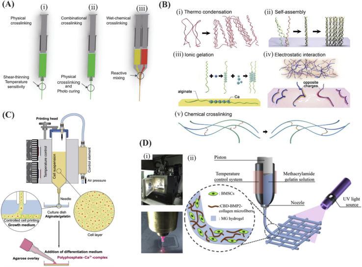
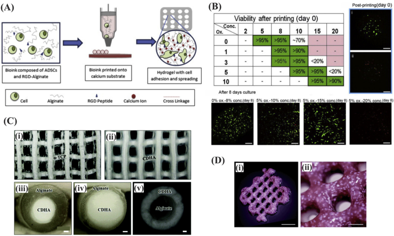
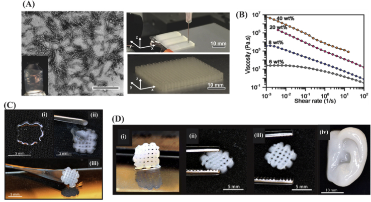
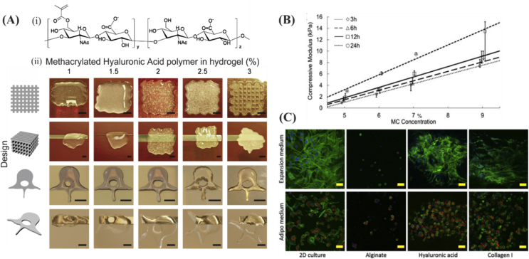
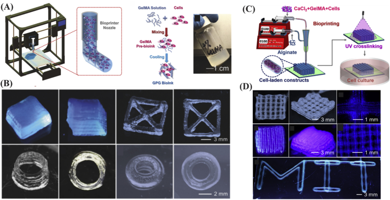
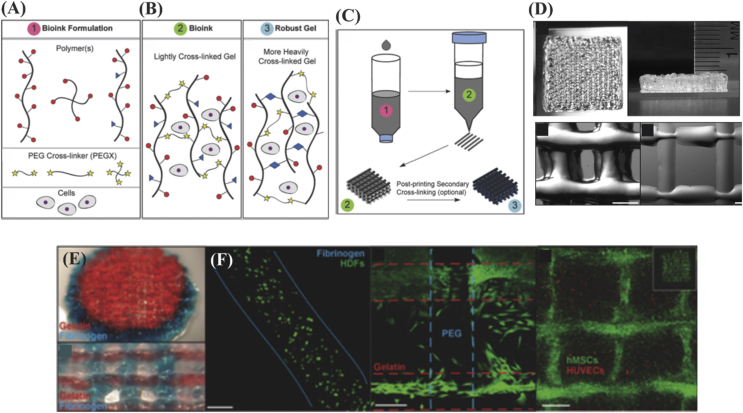
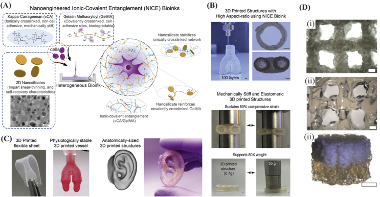
The native tissues are complex structures consisting of different cell types, extracellular matrix materials, and biomolecules. Traditional tissue engineering strategies have not been able to fully reproduce biomimetic and heterogeneous tissue constructs because of the lack of appropriate biomaterials and technologies. However, recently developed three-dimensional bioprinting techniques can be leveraged to produce biomimetic and complex tissue structures. To achieve this, multicomponent bioinks composed of multiple biomaterials (natural, synthetic, or hybrid natural-synthetic biomaterials), different types of cells, and soluble factors have been developed. In addition, advanced bioprinting technologies have enabled us to print multimaterial bioinks with spatial and microscale resolution in a rapid and continuous manner, aiming to reproduce the complex architecture of the native tissues. This review highlights important advances in heterogeneous bioinks and bioprinting technologies to fabricate biomimetic tissue constructs. Opportunities and challenges to further accelerate this research area are also described.

Keywords: Biofabrication; Regenerative medicine; Three-dimensional bioprinting; Tissue engineering.


Figures

Fig. 1
Fig. 1
Schematic representation of the procedure for design and biofabrication of tissue constructs from bioinks mimicking the native tissue parts of figure are reproduced from Zhang et al. with permission from Elsevier.
Fig. 2
Fig. 2
Different crosslinking methods of bioinks. (A) Schematic of crosslinking approaches including ​(i-ii) physical, combinational, and (iii) wet-chemical crosslinking approaches in extrusion printing. Reproduced from Malda et al. with permission from Wiley-VCH Verlag GmbH & Co. (B) Hydrogel crosslinking approaches. (i to iv) Physical crosslinking approaches including: (i) thermally induced polymer chain entanglement, (ii) molecular self-assembly, (iii) ionic gelation, (iv) electrostatic interaction, and (v) chemical crosslinking. (Scale bar ​= ​100 ​μm). Reprinted from Zhang and Khademhosseini , with permission from AAAS. (C) Schematic indicating 3D cell bioprinting of SaOS-2 ​cells in gelatin/alginate. The bioprinted bioink was then put in a calcium chloride (CaCl2) solution. This construct was then covered with an agarose layer and cultured in osteogenic differentiation medium. Reproduced from Neufurth et al. with permission from Elsevier. (D) Schematic indicating the construct photocrosslinking. Reproduced from Du et al. with permission from IOP. BMSCs, bone marrow–derived mesenchymal stem cells; BMP2, bone morphogenetic protein 2.
Fig. 3
Fig. 3
(A) Schematic illustration of droplet-based fabrication process for lattice structure made of human adipose tissue–derived stem cells encapsulated in alginate and (B) the resulting cell viability as a function of bioink parameters. Alginates with medium viscosity resulted in higher cell viability owing to limited nutrition transfer at high and low concentrations. Reproduced from Jia et al. with permission from Elsevier. (C) Representation of 3D printed alpha tricalcium phosphate (α-TCP)/alginate core/shell scaffolds (i) before and (ii) after crosslinking, (iii-iv) fiber cross sections demonstrating the core/shell structure of alginate and calcium-deficient HAp. Scale bar ​= ​100 ​μm. Reproduced from Raja et al. with permission from the Royal Society of Chemistry. (D) Three-dimensional printing of a porous scaffold consisting of alginate, mesenchymal stem cells and, calcium phosphate particles using extrusion printing. Scale bar ​= ​500 ​μm. Reproduced from Loozen et al. with permission from the Royal Society of Chemistry. Hap, hydroxyapatite; RGD, Arg-Gly-Asp.
Fig. 4
Fig. 4
(A) Wood pulp CNC distribution in the aqueous inks (scale bar ​= ​500 ​nm) and photograph of the printed cellular constructs based on CNC inks. (B) Shear-thinning was induced to the ink slightly at 1% CNC concentration and increased significantly at around 10–20% as the viscosity decreased with shear rate. Reproduced from Siqueira et al. with permission from Wiley-VCH Verlag GmbH & Co. (C) The printed constructs for a grid design obtained by using (i) 3% alginate, (ii) 2.5% NFC, or (iii) alginate/NFC inks. Combining the inks resulted in successful and structurally integrated (fully cured) constructs. (D) (i-iii) Mechanical recovery of the meshes and (iv) human ear model obtained by the use of alginate/NFC inks. Reprinted from Markstedt with permission from the American Chemical Society. CNC, cellulose nanocrystals; NFC, nanofibrillated cellulose.
Fig. 5
Fig. 5
Characteristics of printed constructs based on HA modified inks. (A) (i) Chemical structure of methacrylated HA obtained by reacting HA with methacrylic anhydride in an aqueous environment and (ii) printability of methacrylated hyaluronic acid (MeHA) at different concentrations (scale bar ​= ​500 ​μm). The best printability was attained at MeHA concentration of 3%. Reproduced from Poldervaart et al. (B) Compressive elastic properties of HA methylcellulose as a function of methylcellulose at different time points. Reproduced from Law et al. with permission from Elsevier. (C) Comparing cell response in two different culture media (expansion and adipo), for cells seeded on either 2D surface or encapsulated in alginate, HA, or in collagen gels. Promoted cell differentiation and proliferation were seen in HA and collagen I gels (scale bar ​= ​50 ​μm; nuclei, actin filaments, and lipid droplets were shown in blue, green, and red, respectively). Reproduced from Henriksson et al. with permission from IOP Publishing Copyright 2016. HA, hyaluronan.
Fig. 6
Fig. 6
(A) 3D bioprinting of bioinks composed of cells and GelMA. (B) Low concentration 3D GelMA structures were fabricated by taking advantage of shear-thinning properties of GelMA, i.e. cooling down the structures to maintain their structural integrity. Reproduced from Liu et al. with permission from WILEY-VCH Verlag GmbH & Co. (C) Illustration of GelMA/alginate microfibers with core/sheath architecture forming bioprinted constructs through extrusion 3D bioprinting. (D) Alginate sheath allows printing of 3D structures using low concentration GelMA (lower than 2%). Reproduced from Liu et al. with permission from IOP Publishing. GelMA, gelatin methacryloyl.
Fig. 7
Fig. 7
(A) Different types of polymer chain, crosslinker, and cells for developing cell-incorporated inks based on PEG. (B) Ink configuration after lightly and heavy crosslinking of PEG. (C) Three-dimensional printing strategy of developed PEG ink. Secondary crosslinking may be applied for heavily crosslinking the polymer chains after the 3D printing process was completed. (D) Photograph of 3D printed structures using PEG-gelatin bioinks (scale bar ​= ​500 ​μm). (E) Examples of combined 3D bioprinting of PEG-gelatin (red) and PEG-fibrinogen (blue) bioinks in spheroidal and grid designs and (F) cell viability results associated with using 3 w/v% fibrinogen in PEG, PEG-PEG, and PEG-gelatin (scale bar ​= ​200 ​μm). Human umbilical vein endothelial cells (HUVECs) seeded with human MSCs, which filled the pore spaces in the internal structure. Reproduced from Rutz et al. with permission from WILEY-VCH Verlag GmbH & Co. PEG, polyethylene glycol; MSC, mesenchymal stem cell.
Fig. 8
Fig. 8
(A) Nanoengineered ionic-covalent entanglement (NICE) bioinks developed by taking advantage of nanoparticle ingredients: (1) Kappa-Carrageenan (κCA) for ionic crosslinking, (2) GelMA for covalent crosslinking, tissue adhesion, and biodegradability, and (3) 2D nanosilicates for having shear-thinning properties. (B) The NICE-based printed constructs exhibit promising mechanical recovery behavior (scale bar ​= ​1 ​mm). (C) High printing fidelity was achieved by the NICE ink because of versatile printing of complex 3D structures and human organs. Reprinted with permission from Ref. . Copyright 2018 American Chemical Society. (D) Porous constructs fabricated by gelatin methacrylamide-gellan gum MSC-laden bioinks: (i) MSC-laden layer (scale bar ​= ​400 ​μm), (ii) GelMA-gellan gum layer (scale bar ​= ​400 ​μm), and (iii) perspective photograph of the bilayered GelMA-gellan gum cylindrical osteochondral graft model (scale bar ​= ​4 ​mm). Reproduced from Levato et al. with permission from IOP Publishing. GelMA, gelatin methacryloyl, MSC, mesenchymal stem cell.
Fig. 9
Fig. 9
(A) Schematic illustration of reversible gelation of the shear-thinning bioinks consisting of κCA and nSi. Double helical structure was formed with controlling temperature and the structure can ionically crosslink through K+ ions. This allowed the 3D printing of highly complex structures. Reprinted from Wilson et al. with permission from the American Chemical Society. (B) Representation of the fabrication scheme of (i) cell-laden collagen scaffold (CLCS), (ii) cell-laden α-TCP/collagen fabricated through cell dipping process (TC-CDIP), and (iii) cell-laden α-TCP/collagen fabricated through cell printing process (TC-CPRINT). (C) Comparing the osteogenic activity of collagen vs. α-TCP/collagen scaffolds in terms of (i) ALP activity, (ii) relative calcium deposition, and (iii) osteopontin (OPN). Reproduced from Kim et al. . ALP, alkaline phosphatase.
Fig. 10
Fig. 10
Scalable process of ink development by combining elastomer solution with graphene for fabricating porous and conductive scaffolds for tissue engineering applications. Reprinted from Jakus et al. with permission from the American Chemical Society.
Fig. 11
Fig. 11
Schematic illustration of different 3D printing systems that have been used to produce multimaterial constructs.
Fig. 12
Fig. 12
Three-dimensional printing with (A) single and (B, C, and D) multiprint head systems . PCL, poly(ε-caprolactone); dECM, decellularized ECM

References

    1. Ashammakhi N., Hasan A., Kaarela O., Byambaa B., Sheikhi A., Gaharwar A.K. Advancing frontiers in bone bioprinting. Adv. Healthc. Mater. 2019;8(7):e1801048. - PubMed
    1. Jakus A.E., Rutz A.L., Shah R.N. Advancing the field of 3D biomaterial printing. Biomed. Mater. 2016;11(1):014102. - PubMed
    1. McBeth C., Lauer J., Ottersbach M., Campbell J., Sharon A., Sauer-Budge A.F. 3D bioprinting of GelMA scaffolds triggers mineral deposition by primary human osteoblasts. Biofabrication. 2017;9(1):015009. - PubMed
    1. Kang H.-W., Lee S.J., Ko I.K., Kengla C., Yoo J.J., Atala A. A 3D bioprinting system to produce human-scale tissue constructs with structural integrity. Nat. Biotechnol. 2016;34(3):312. - PubMed
    1. Murphy S.V., Atala A. 3D bioprinting of tissues and organs. Nat. Biotechnol. 2014;32(8):773. - PubMed

LinkOut - more resources