. 2019 Apr 20:2:100005.
doi: 10.1016/j.mtbio.2019.100005. eCollection 2019 Mar.

Biomimetic hydroxyapatite/collagen composite drives bone niche recapitulation in a rabbit orthotopic model

Affiliations

Biomimetic hydroxyapatite/collagen composite drives bone niche recapitulation in a rabbit orthotopic model

S Minardi et al. Mater Today Bio. .

Abstract

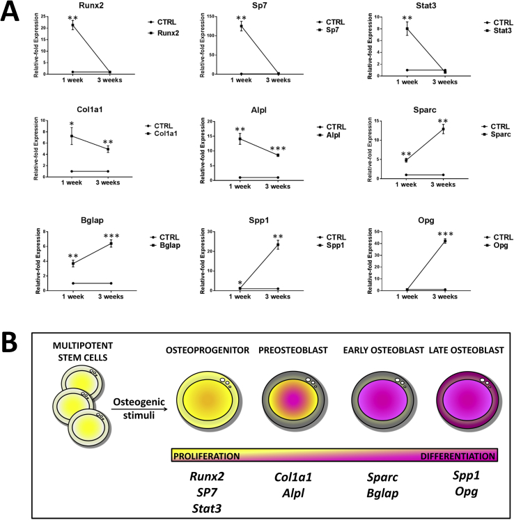
Synthetic osteoinductive materials that mimic the human osteogenic niche have emerged as ideal candidates to address this area of unmet clinical need. In this study, we evaluated the osteoinductive potential in a rabbit orthotopic model of a magnesium-doped hydroxyapatite/type I collagen ​(MHA/Coll) composite. The composite was fabricated to exhibit a highly fibrous structure of carbonated MHA with 70% (±2.1) porosity and a Ca/P ratio of 1.5 (±0.03) as well as a diverse range of elasticity separated to two distinct stiffness peaks of low (2.35 ​± ​1.16 ​MPa) and higher (9.52 ​± ​2.10 ​MPa) Young's Modulus. Data suggested that these specific compositional and nanomechanical material properties induced the deposition of de novo mineral phase, while modulating the expression of early and late osteogenic marker genes, in a 3D in vitro model using human bone marrow-derived mesenchymal stem cells (hBM-MSCs). When tested in the rabbit orthotopic model, MHA/Col1 scaffold induction of new trabecular bone mass was observed by DynaCT scan, only 2 weeks after implantation. Bone histomorphometry at 6 weeks revealed a significant amount of de novo bone matrix formation. qPCR demonstrated MHA/Coll scaffold full cellularization in vivo and the expression of both osteogenesis-associated genes (Spp1, Sparc, Col1a1, Runx2, Dlx5) as well as hematopoietic (Vcam1, Cd38, Sele, Kdr) and bone marrow stromal cell marker genes (Vim, Itgb1, Alcam). Altogether, these data provide ​evidence of the solid osteoinductive potential of MHA/Coll and its suitability for multiple approaches of bone regeneration.

Keywords: Biomimetic material; Bone regeneration; Collagen; Hydroxyapatite; Stem cell.


Figures

Fig. 1
Fig. 1
(A) Representative TGA analysis of the mineral content of MHA/Coll and (B) Young's Modulus measurements. TGA, thermal gravimetric analysis; MHA/Coll, magnesium-doped hydroxyapatite/type I collagen.
Fig. 2
Fig. 2
Confocal laser microscopy and SEM images of hBM-MSCs (green) cultured on Coll, Coll in osteogenic media and MHA/Coll, at 3 weeks (A-C ​and D-F, respectively). Relative amount (intensity) of magnesium, phosphate and calcium in the scaffolds, for the three corresponding experimental groups, quantified by EDS analysis. Data are normalized to cell-free Coll and MHA/Coll scaffolds, as baselines. SEM, scanning electron microscopy; hBM-MSC, human bone marrow-derived mesenchymal stem cell; MHA/Coll, magnesium-doped hydroxyapatite/type I collagen.
Fig. 3
Fig. 3
(A) Relative-fold expression of osteoprogenitor, preosteoblast, and early and late osteoblast marker genes at 1week and 3 weeks of in vitro culture of hBM-MSCs cultured on MHA/Coll and control Coll scaffolds. Results were first normalized to the expression of the housekeeping gene GAPDH ​and then expressed as “relative-fold expression” in comparison to the expression level of the same gene from the CTRL (Coll). Values are reported as mean ​± ​standard deviation. A value of p ​< ​0.05 was considered statistically significant: *p ​< ​0.05, **p ​< ​0.01, ***p ​< ​0.001. (B) Schematic representing the different stages of osteogenic differentiation of mesenchymal stem cells, with associated relevant marker genes. hBM-MSC, human bone marrow-derived mesenchymal stem cell; MHA/Coll, magnesium-doped hydroxyapatite/type I collagen.
Fig. 4
Fig. 4
Relative-fold expression of MAGT1 at 1week and 3 weeks of in vitro culture of hBM-MSCs cultured on MHA/Coll and Coll (CTRL). TRPM7 was not found expressed at neither time points (data not shown). Results were first normalized to the expression of the housekeeping gene GAPDH ​and then expressed as “relative-fold expression” in comparison to the expression level of the same gene from the CTRL. Values are reported as mean ​± ​standard deviation. A value of p ​< ​0.05 was considered statistically significant: *p ​< ​0.05, **p ​< ​0.01. hBM-MSC, human bone marrow-derived mesenchymal stem cell; MHA/Coll, magnesium-doped hydroxyapatite/type I collagen; MAGT1, magnesium transporter 1.
Fig. 5
Fig. 5
3D rendering obtained by DynaCT scan of a representative spine showing radiologic bone mass formation in correspondence of the scaffold (arrowhead) (A, B). Bone-like scaffold at 6 weeks, upon retrieval, showing integration with surrounding tissue (arrowhead) (C). MHA/Coll allowed early and robust trabecular bone formation as well as cortical bone ingrowth as early as 2 weeks (D).
Fig. 6
Fig. 6
Von Kossa (A, B), Goldner's Trichrome (C, D), and hematossilin-eosin (E, F) staining of non-demineralized vertebral trabecular bone and MHA/Coll specimens at 6 weeks, respectively. TB, trabecular bone; OS, osteoid; MP, mineral phase; OB, osteoblast; OC, osteoclast; BMSC, bone marrow stem cell.
Fig. 7
Fig. 7
Releative-fold Expression of osteogenic (A), hematopoitic (B), and bone marrow–associated marker genes (C) detected for MHA/Coll group at 6 weeks after implantation in the orthotopic model in rabbit, compared with naïve spinal bone. Results were first normalized to the expression of the housekeeping gene GAPDH ​and then expressed as “relative-fold expression” in comparison to the expression level of the same gene from the CTRL. Values are reported as mean ​± ​standard deviation. A value of p ​< ​0.05 was considered statistically significant: *p ​< ​0.05, **p ​< ​0.01, ***p ​< ​0.001. TP, transverse process; MHA/Coll, magnesium-doped hydroxyapatite/type I collagen.

References

    1. Schubert T., Lafont S., Beaurin G., Grisay G., Behets C., Gianello P., Dufrane D. Critical size bone defect reconstruction by an autologous 3D osteogenic-like tissue derived from differentiated adipose MSCs. Biomaterials. 2013;34(18):4428–4438. - PubMed
    1. Arrington E.D., Smith W.J., Chambers H.G., Bucknell A.L., Davino N.A. Complications of iliac crest bone graft harvesting. Clin. Orthop. Relat. Res. 1996;329:300–309. - PubMed
    1. Bucholz R.W. Nonallograft osteoconductive bone graft substitutes. Clin. Orthop. Relat. Res. 2002;395:44–52. - PubMed
    1. Silber J.S., Anderson D.G., Daffner S.D., Brislin B.T., Leland J.M., Hilibrand A.S., Vaccaro A.R., Albert T.J. Donor site morbidity after anterior iliac crest bone harvest for single-level anterior cervical discectomy and fusion. Spine. 2003;28(2):134–139. - PubMed
    1. Campana V., Milano G., Pagano E., Barba M., Cicione C., Salonna G., Lattanzi W., Logroscino G. Bone substitutes in orthopaedic surgery: from basic science to clinical practice. J. Mater. Sci. Mater. Med. 2014;25(10):2445–2461. - PMC - PubMed