

Shape-defined solid micro-objects from poly(d,l-lactic acid) as cell-supportive counterparts in bottom-up tissue engineering
- PMID: 32159154
- PMCID: PMC7061620
- DOI: 10.1016/j.mtbio.2019.100025
Shape-defined solid micro-objects from poly(d,l-lactic acid) as cell-supportive counterparts in bottom-up tissue engineering
Abstract
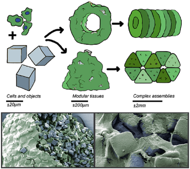
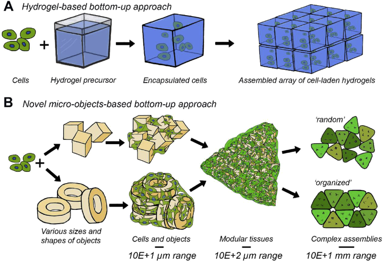
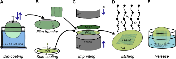
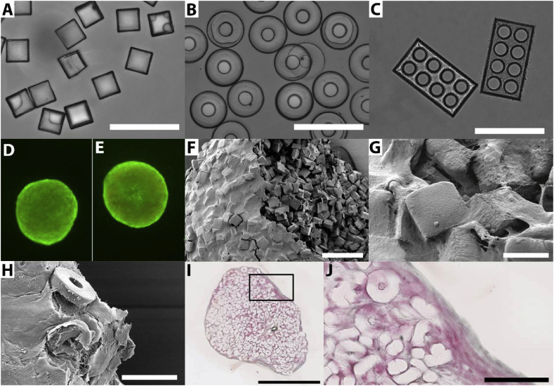
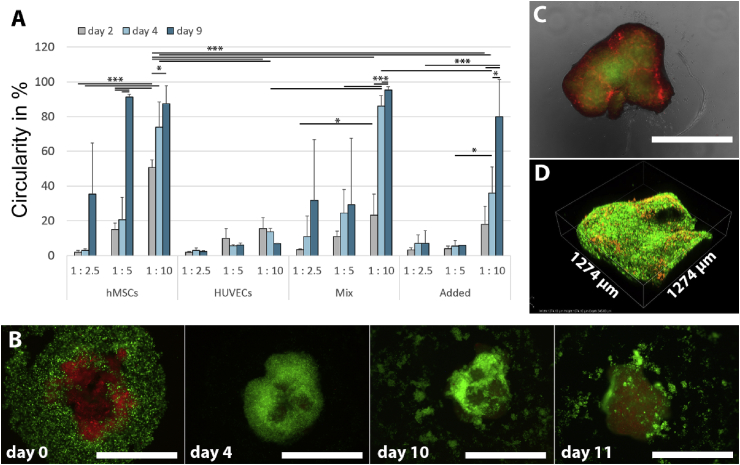
In bottom-up tissue engineering, small modular units of cells and biomaterials are assembled toward larger and more complex ones. In conjunction with a new implementation of this approach, a novel method to fabricate microscale objects from biopolymers by thermal imprinting on water-soluble sacrificial layers is presented. By this means, geometrically well-defined objects could be obtained without involving toxic agents in the form of photoinitiators. The micro-objects were used as cell-adhesive substrates and cell spacers in engineered tissues created by cell-guided assembly of the objects. Such constructs can be applied both for in vitro studies and clinical treatments. Clinically relevantly sized aggregates comprised of cells and micro-objects retained their viability up to 2 weeks of culture. The aggregation behavior of cells and objects showed to depend on the type and number of cells applied. To demonstrate the micro-objects' potential for engineering vascularized tissues, small aggregates of human bone marrow stromal cells (hMSCs) and micro-objects were coated with a layer of human umbilical vein endothelial cells (HUVECs) and fused into larger tissue constructs, resulting in HUVEC-rich regions at the aggregates' interfaces. This three-dimensional network-type spatial cellular organization could foster the establishment of (premature) vascular structures as a vital prerequisite of, for example, bottom-up-engineered bone-like tissue.
Keywords: Bone tissue engineering; Hot embossing/thermal imprinting; Human bone marrow stromal cells; Human umbilical vein endothelial cells; Poly(lactic acid); Self-assembly.
© 2019 The Authors.
Figures
Similar articles
-
Engineered micro-objects as scaffolding elements in cellular building blocks for bottom-up tissue engineering approaches.Adv Mater. 2014 Apr 23;26(16):2592-9. doi: 10.1002/adma.201304539. Epub 2014 Jan 7. Adv Mater. 2014. PMID: 24395427
-
Co-culture of human umbilical vein endothelial cells and human bone marrow stromal cells into a micro-cavitary gelatin-methacrylate hydrogel system to enhance angiogenesis.Mater Sci Eng C Mater Biol Appl. 2019 Sep;102:906-916. doi: 10.1016/j.msec.2019.04.089. Epub 2019 Apr 29. Mater Sci Eng C Mater Biol Appl. 2019. PMID: 31147062
-
Polymeric Microspheres/Cells/Extracellular Matrix Constructs Produced by Auto-Assembly for Bone Modular Tissue Engineering.Int J Mol Sci. 2021 Jul 23;22(15):7897. doi: 10.3390/ijms22157897. Int J Mol Sci. 2021. PMID: 34360672 Free PMC article.
-
Coupling Osteogenesis and Vasculogenesis in Engineered Orthopedic Tissues.Tissue Eng Part B Rev. 2021 Jun;27(3):199-214. doi: 10.1089/ten.TEB.2020.0132. Epub 2020 Sep 25. Tissue Eng Part B Rev. 2021. PMID: 32854589 Free PMC article. Review.
-
Modular Tissue Assembly Strategies for Biofabrication of Engineered Cartilage.Ann Biomed Eng. 2017 Jan;45(1):100-114. doi: 10.1007/s10439-016-1609-3. Epub 2016 Apr 12. Ann Biomed Eng. 2017. PMID: 27073109 Review.
Cited by
-
Modular Orthopaedic Tissue Engineering With Implantable Microcarriers and Canine Adipose-Derived Mesenchymal Stromal Cells.Front Bioeng Biotechnol. 2020 Jul 22;8:816. doi: 10.3389/fbioe.2020.00816. eCollection 2020. Front Bioeng Biotechnol. 2020. PMID: 32775324 Free PMC article.
-
Chips for Biomaterials and Biomaterials for Chips: Recent Advances at the Interface between Microfabrication and Biomaterials Research.Adv Healthc Mater. 2021 Jul;10(14):e2100371. doi: 10.1002/adhm.202100371. Epub 2021 May 25. Adv Healthc Mater. 2021. PMID: 34033239 Free PMC article. Review.
-
Hollow pollen grains as scaffolding building blocks in bone tissue engineering.Bioimpacts. 2022;12(3):183-193. doi: 10.34172/bi.2021.24. Epub 2021 Dec 18. Bioimpacts. 2022. PMID: 35677670 Free PMC article.
-
Nanofunctionalized Microparticles for Glucose Delivery in Three-Dimensional Cell Assemblies.ACS Appl Mater Interfaces. 2024 Apr 10;16(14):17347-17360. doi: 10.1021/acsami.4c02321. Epub 2024 Apr 1. ACS Appl Mater Interfaces. 2024. PMID: 38561903 Free PMC article.
-
Engineering Heterogeneous Tumor Models for Biomedical Applications.Adv Sci (Weinh). 2024 Jan;11(1):e2304160. doi: 10.1002/advs.202304160. Epub 2023 Nov 9. Adv Sci (Weinh). 2024. PMID: 37946674 Free PMC article. Review.
References
-
- Langer R., Vacanti J.P. Tissue engineering. Science. 1993;260(5110):920–926. - PubMed
-
- Marx V. Tissue engineering: organs from the lab. Nature. 2015;522(7556):373–377. - PubMed
-
- Moroni L. Chapter 1 – Tissue engineering: an introduction. In: van Blitterswijk C., editor. Tissue Engineering. Academic Press; 2014. pp. 1–21.
LinkOut - more resources
Full Text Sources
