. 2020 Jul;237(1):176-187.
doi: 10.1111/joa.13181. Epub 2020 Mar 11.

The incudopetrosal joint of the human middle ear: a transient morphology in fetuses

Affiliations

The incudopetrosal joint of the human middle ear: a transient morphology in fetuses

José Francisco Rodríguez-Vázquez et al. J Anat. 2020 Jul.

Abstract

In spite of the amount of research on fetal development of the human middle ear and ear ossicles, there has been no report showing a joint between the short limb of incus and the otic capsule or petrous part of the temporal bone. According to observations of serial histological sections from 65 embryos and fetuses at 7-17 weeks of development, the incudopetrosal joint exhibited a developmental sequence similar to the other joints of ossicles, with an appearance of an interzone followed by a trilaminar configuration at 7-12 weeks, a joint cavitation at 13-15 weeks and development of intraarticular and capsular ligaments at 16-17 weeks. These processes occurred at the same time or slightly later than any other joint. Thus, the joint development might coordinate with vibrating ossicles in utero. The growing short limb of incus appeared to accelerate an expansion of the epitympanic recess of the tympanic cavity. Additional observations of five late-stage fetuses demonstrated the incudopetrosal joint located in the fossa incudis joint changing to syndesmosis. Consequently, a real joint with a cavity existed transiently between the human neurocranium and the first pharyngeal arch derivative (i.e. incus) in contrast to the tympanostapedial joint or syndesmosis between the neurocranium and the second arch derivative. The newly described joint might have an effect on the widely accepted primary jaw concept: the mammalian jaw should thus have been created within the first pharyngeal arch, although the connection with neurocranium by the stapes is of a different origin.

Keywords: human fetus; incudopetrosal joint; incus; middle ear; otic capsule.


Figures

Figure 1
Figure 1
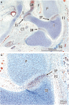
Precartilaginous union of incus‐otic capsule. (a,b) 7 WD. Human embryo of 20 mm GL (20 CS). Frontal section. HE staining. (b) Enlargement of (a). Lateral to the facial nerve (F) and close to the semicircular duct (LS), a precartilaginous connection: union of incus‐otic capsule (IP) is distinguishable between the short limb of the incus (SI) and the lateral protrusion of the otic capsule (P). The initial perichondrium (arrows) and precartilaginous cells are continuous in the union of the incus‐otic capsule. (c,d) 7 WD. Human embryo of 23 mm GL (21 CS). Horizontal section. HE staining. (d) Enlargement of (c). The union between the incus and otic capsule (IP) is characterized by the continuity of the precartilaginous cells and the primordium of perichondrium (arrows). (e,f) 7 WD. Human embryo of 23.5 mm GL (21 CS). Horizontal section. HE staining. (f) Enlargement of (e). Topographical anatomy of the incus (I) and malleus (M). Union between the incus and otic capsule (IP) and the incudomalleolar joint (IM) was observed. In the incus‐otic capsule union, precartilaginous cells and perichondral sheath (arrows) are continuous. Lateral to the incus, a mesenchymal condensation was evident, being the origin of the squamous part of the temporal bone (SQ). For other abbreviations, see common abbreviations below. AP, articular apophysis; AS, articular surface; BI, body of incus; C, articular capsule of the incudopetrosal joint; CA, articular cavity of the incudopetrosal joint; CT, chorda tympani nerve; D, disk of the temporomandibular joint; DM, discomalleolar ligament; E, epitympanic recess; EE, external ear; F, facial nerve; FI, fossa incudis; G, gonial/anterior process of malleus; GG, geniculate ganglion; H, hyoid artery; I, incus; IE, internal ear; IL, intraarticular/interosseous ligament; IM, incudomalleolar joint; IS, incudostapedial joint; IP, incudopetrosal joint/ union incus‐otic capsule; LL, long limb of incus; LS, lateral semicircular canal; M, malleus; MK, Meckel´s cartilage; P, petrous part of temporal bone/ otic capsule; PG, parotid gland; PL, posterior ligament of incus; PS, petrosquamosal fissure; R, Reichert´s cartilage; S, stapes; SA, stapedial artery; SM, stapedial muscle; SQ, squamous part of temporal bone; SI, short limb of incus; SV, synovial of the incudopetrosal joint; TB, tympanic bone; TM, temporalis muscle; TS, tympanostapedial joint; TT, tegmen tympani
Figure 2
Figure 2
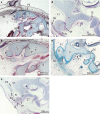
Cartilaginous union between the incus and otic capsule with the anlage of the fossa incudis. (a,b) 8 WD. Human embryo of 26.5 mm GL (22 CS). Horizontal section. Azan staining. (b) Enlargement of (a). There is a cartilaginous continuity in the incus‐otic capsule (IP). The perichondral sheath of incus and otic capsule is continuous (arrows). (c,d) 8 WD. Human embryo of 29 mm GL (23 CS). Horizontal section. HE staining. (d) Enlargement of (c). A cartilaginous continuity in the incus‐otic capsule (IP) and incudomalleolar joint (IM) is seen. The perichondral layer of incus and otic capsule is continuous (arrows) without interruption. (e,f) 8 WD. Human embryo of 28 mm GL (23 CS). Frontal section. Azan staining. (f) Enlargement of (e). The short limb of the incus (SI) is surrounded by the otic capsule (P; petrous part) and the anlage of the squamous part of the temporal bone (SQ). A small cavity appears in the posterior wall of the tympanic cavity, i.e. the future fossa incudis (FI). For other abbreviations, see Figure 1
Figure 3
Figure 3
Homogeneous interzone phase of the incudopetrosal joint in the fossa incudis. (a,b) 9 WD. Human fetus of 45 mm GL. Horizontal section. Azan staining. (b) Enlargement of (a). A homogeneous interzone is observed in the incudopetrosal joint (IP) between the dorsal end of the short limb of the incus (SI) and the otic capsule (P). The tympanostapedial joint (IS) is in the interzone phase, and the cavity of incudomalleolar joint (IM) has become larger. For other abbreviations, see Figure 1
Figure 4
Figure 4
Establishment of the petrosquamosal fissure and closure of the posterior walls of the tympanic cavity. (a,b) 10 WD. Human fetus of 52 mm GL. Horizontal section. Azan staining. (b) Enlargement of (a). The petrosquamosal fissure (PS) has formed between the squamous part (SQ) and the petrous part (P, otic capsule) of the temporal bone. (c,d) 11 WD. Human fetus of 64 mm GL. Frontal sections. HE staining. (d) Enlargement of (c). The tegmen tympani (TT) has closed the upper and posterior walls of the tympanic cavity. The tegmen tympani (TT) has formed a wide petrosquamosal fissure (PS) with the squamous part of the temporal bone (SQ). The short limb of the incus (SI) is located in a recess, i.e. the future fossa incudis (FI). The otic capsule (P) has an eminence or protrusion where the interzone of the incudopetrosal joint (IP) is located. The posterior ligament of the incus (PL) has begun to develop as a dense mesenchyme between the squamous part of the temporal bone (SQ) and short limb of the incus (SI). (e) 12 WD. Human fetus of 74.5 mm GL. Frontal section. Azan staining. The incudopetrosal joint (IP) located in the fossa incudis (FI) is in the trilaminar interzone phase. For other abbreviations, see Figure 1
Figure 5
Figure 5
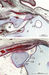
Cavitation phase of the incudopetrosal joint. (a) 13 WD. Human fetus of 105 mm GL. Horizontal section. Azan staining. The topographical anatomy of the tympanic cavity is observed at the level of the fossa incudis (FI), as well as the location and relationships of the incudopetrosal joint (IP) in the initial cavitation phase. (b) 14 WD. Human fetus of 120 mm GL. Horizontal section. Azan staining. The cavity (CA) of the incudopetrosal joint (IP) is wider and delimited by the initial capsule (C) and synovial membrane has appeared (SV). The anlage of the posterior ligament of the incus (PL) is visible between the short limb of the incus (SI) and the squamous part of the temporal bone (SQ). For other abbreviations, see Figure 1
Figure 6
Figure 6
Change of the incudopetrosal joint to a syndesmosis. (a, b) 16 WD. Human fetus of 144 mm GL. Horizontal section. OF staining. (b) Enlargement of (a). Endochondral ossification has begun in the petrous part of the temporal bone (P) near the facial nerve (F) but has not reached the short limb of the incus (SI). The cavity of the incudopetrosal joint (IP) is reduced in size and located in the fossa incudis (FI) in the posterior wall of the epitympanic recess (e). (c) 17 WD. Human fetus of 150 mm. Horizontal section. HE staining. Development of the intraarticular and capsular ligament (IL) has begun. For other abbreviations, see Figure 1
Figure 7
Figure 7
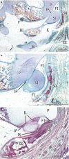
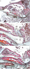
Incudopetrosal joint in a 25 WD human fetus. (a‐c) Human fetus of 240 mm GL. Sagittal sections. Azan staining. (a) The most lateral section. (c) The most medial section. (b) Enlargement of (a). Topographical anatomy of the tympanic cavity. The incudomalleolar (IM) and incudopetrosal (IP) joints can be observed. The incudopetrosal joint (IP) has changed to a syndesmosis, an intrinsic interosseous ligament (IL) joins both articular surfaces, and an extrinsic ligament, the posterior ligament (PL) of the incus, has joined the lateral side of the end of the short limb of the incus (SI) to the squamous part of the temporal bone (SQ). * Posterior tympanic isthmus. For other abbreviations, see Figure 1
Figure 8
Figure 8
Incudopetrosal joint in a 34 WD human fetus. (a‐c) Human fetus of 340 mm. Horizontal sections. Azan staining. (a) The most superior section. (c) The most inferior section. (c) Enlargement of (b). Topographical anatomy of the incudopetrosal joint (IP) at the tympanic cavity. The incudopetrosal joint (IP) is located in the fossa incudis (FI) in the angle or recess that forms the squamous part (SQ) and the petrous parts (P) of the temporal bone, in the posterior wall of the tympanic cavity at the epitympanic recess lateral to the protrusion of the facial canal and facial nerve (F), and inferior and medial to the lateral semicircular canal (LS). * Posterior tympanic isthmus. For other abbreviations, see Figure 1
Figure 9
Figure 9
Diagrammatic representation of the steps of development of the incudopetrosal joint (IP). (a) Precartilaginous union incus‐otic capsule (7WD). (b) Cartilaginous union between the incus and the otic capsule (8 WD). (c) Homogeneous interzone phase (9–11 WD). (d) Trilaminar interzone phase (12 WD). (e) Cavitation phase (13–15 WD). (f) Joint syndesmosis phase (17–34 WD). For other abbreviations, see Figure 1
Figure 10
Figure 10
Lateral view diagrammatic representation of the incudopetrosal joint and the ossicles of the middle ear in midterm human fetuses. The incus (I) connects with the neurocranium (P, petrous part of the temporal bone) via the incudopetrosal joint (IP). The derivatives of the first arc (I, incus) are colored light brown, and the cartilage of the first pharyngeal arch (MK, Meckel´s cartilage) and its derivative malleus (M) dark brown. The derivative of the second pharyngeal arch (S, stapes) is colored light blue, and the cartilage of the second arch (R, Reichert's cartilage) dark blue. The Meckel cartilage (MK) passes through the tympanosquamosal fissure (red circle). The discomalleolar ligament (DM) connects the disk of the temporomandibular joint (D) to the inferior process of the tegmen tympani (TT) and descends into the fissure to close it (Rodríguez‐Vázquez et al., 2011). For other abbreviations, see Figure 1

Similar articles

Cited by

References

    1. Aimi, K. (1971) The clinical significance of epitympanic mucosal folds. Archives of Otolaryngology, 94, 499–508. - PubMed
    1. Aimi, K. (1978) The tympanic isthmus: its anatomy and clinical significance. Laryngoscope, 88, 1067–1081. - PubMed
    1. Anson, B.J. and Donaldson, J.A. (1973) Surgical anatomy of the temporal bone and ear, 2nd edition Philadelphia: W.B. Saunders Company.
    1. Anson, B.J. , Hanson, J.R. and Richany, S.F. (1960) Early embryology of the auditory ossicles and associated structures in relation to certain anomalies observed clinically. Annals of Otology, Rhinology, and Laryngology, 69, 427–447. - PubMed
    1. Ars, B. (1989) Organogenesis of the middle ear structures. Journal of Laryngology and Otology, 103, 16–21. - PubMed