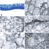
Evidence of autophagic vesicles in a patient with Lisch corneal dystrophy
- PMID: 32159595
- PMCID: PMC11826730
- DOI: 10.5935/0004-2749.20200027
Evidence of autophagic vesicles in a patient with Lisch corneal dystrophy
Abstract
Lisch corneal dystrophy is a rare corneal disease characterized by the distinctive feature of highly vacuolated cells. Although this feature is important, the nature of these vacuoles within corneal cells remains unknown. Here, we sought to analyze corneal cells from a patient diagnosed with Lisch dystrophy to characterize the vacuoles within these cells. Analyses using histopathology examination, confocal microscopy, and transmission electron microscopy were all consistent with previous descriptions of Lisch cells. Importantly, the vacuoles within these cells appeared to be autophagosomes and autolysosomes, and could be stained with an anti-microtubule-associated protein 1A/1B-light chain 3 (LC3) antibody. Taken together, these findings indicate that the vacuoles we observed within superficial corneal cells of a patient with Lisch corneal dystrophy constituted autophagosomes and autolysosomes; this finding has not been previously reported and suggests a need for further analyses to define the role of autophagy in this ocular disease.
A distrofia corneana de Lisch é uma doença rara, caracterizada principalmente pela presença de células altamente vacuoladas. Embora esta característica seja importante, a natureza desses vacúolos dentro das células da córnea permanece des conhecida. Aqui, procuramos analisar as células da córnea de um paciente diagnosticado com distrofia de Lisch para caracterizar os vacúolos dentro dessas células. Análises utilizando exame histopatológico, microscopia confocal e microscopia eletrônica de transmissão foram todas consistentes com descrições previas de células de Lisch. Importante, os vacúolos dentro dessas células pareciam ser autofagossomos e autolisossomos, e poderiam ser corados com um anticorpo proteico 1A/1B-cadeia leve 3 (LC3) da proteína anti-microtúbulo associado a microtúbulos. Em conjunto, esses achados indicam que os vacúolos observados nas células superficiais da córnea de um paciente com distrofia corneana de Lisch constituíram autofagossomos e autolisossomos. Esse achado não foi relatado anteriormente e sugere a necessidade de mais análises para definir o papel da autofagia nessa doença ocular.
Conflict of interest statement
Figures


References
-
- Oliver VF, Vincent AL. The genetics and pathophysiology of IC3D category 1 corneal dystrophies: a review. Asia Pac J Ophthalmol (Phila) 2016;5(4):272–281. - PubMed
-
- Weiss JS, Møller HU, Aldave AJ, Seitz B, Bredrup C, Kivelä T, et al. IC3D classification of corneal dystrophies-edition 2. Cornea. 2015;34(2):117–159. - PubMed
-
- Lisch W, Büttner A, Oeffner F, Böddeker I, Engel H, Lisch C, et al. Lisch corneal dystrophy is genetically distinct from Meesmann corneal dystrophy and maps to xp22.3. Am J Ophthalmol. 2000;130(4):461–468. - PubMed
-
- Lisch W, Steuhl KP, Lisch C, Weidle EG, Emmig CT, Cohen KL, et al. A new, band-shaped and whorled microcystic dystrophy of the corneal epithelium. Am J Ophthalmol. 1992;114(1):35–44. - PubMed
Publication types
MeSH terms
Supplementary concepts
LinkOut - more resources
Full Text Sources

